Engineering ESPT Pathways Based on Structural Analysis of LSSmKate Red Fluorescent Proteins with Large Stokes Shift.
Piatkevich, K.D., Malashkevich, V.N., Almo, S.C., Verkhusha, V.V.(2010) J Am Chem Soc 132: 10762-10770
- PubMed: 20681709 Search on PubMedSearch on PubMed Central
- DOI: https://doi.org/10.1021/ja101974k
- Primary Citation Related Structures:
3NT3, 3NT9 - PubMed Abstract:
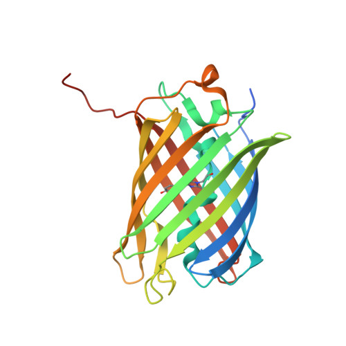
LSSmKate1 and LSSmKate2 are monomeric red fluorescent proteins (RFPs) with large Stokes shifts (LSSs), which allows for efficient separation of absorbance and emission maxima, as well as for excitation with conventional two-photon laser sources. These LSSmKates differ by a single amino acid substitution at position 160 and exhibit absorbance maxima around 460 nm, corresponding to a neutral DsRed-like chromophore. However, excitation at 460 nm leads to fluorescence emission above 600 nm. Structures of LSSmKate1 and LSSmKate2, determined at resolutions of 2.0 and 1.5 A, respectively, revealed that the predominant DsRed-chromophore configurations are cis for LSSmKate1 but trans for LSSmKate2. Crystallographic and mutagenesis analyses, as well as isotope and temperature dependences, suggest that an excited-state proton transfer (ESPT) is responsible for the LSSs observed in LSSmKates. Hydrogen bonding between the chromophore hydroxyl and Glu160 in LSSmKate1 and a proton relay involving the chromophore tyrosine hydroxyl, Ser158, and the Asp160 carboxylate in LSSmKate2 represent the putative ESPT pathways. Comparisons with mKeima LSS RFP suggest that similar proton relays could be engineered in other FPs. Accordingly, we mutated positions 158 and 160 in several conventional red-shifted FPs, including mNeptune, mCherry, mStrawberry, mOrange, and mKO, and the resulting FP variants exhibited LSS fluorescence emission in a wide range of wavelengths from 560 to 640 nm. These data suggest that different chromophores formed by distinct tripeptides in different environments can be rationally modified to yield RFPs with novel photochemical properties.
- Department of Anatomy and Structural Biology, Gruss-Lipper Biophotonics Center, Albert Einstein College of Medicine, 1300 Morris Park Avenue, Bronx, New York 10461, USA.
Organizational Affiliation: