3NOJ | pdb_00003noj

The structure of HMG/CHA aldolase from the protocatechuate degradation pathway of Pseudomonas putida


Experimental Data Snapshot

  • Method: X-RAY DIFFRACTION
  • Resolution: 1.82 Å
  • R-Value Free: 
    0.171 (Depositor), 0.177 (DCC) 
  • R-Value Work: 
    0.159 (Depositor), 0.166 (DCC) 
  • R-Value Observed: 
    0.159 (Depositor) 

Starting Model: experimental
View more details

wwPDB Validation 3D Report Full Report

Validation slider image for 3NOJ

This is version 2.0 of the entry. See complete history

Literature

Structural and Kinetic Characterization of 4-Hydroxy-4-methyl-2-oxoglutarate/4-Carboxy-4-hydroxy-2-oxoadipate Aldolase, a Protocatechuate Degradation Enzyme Evolutionarily Convergent with the HpaI and DmpG Pyruvate Aldolases.

Wang, W.Mazurkewich, S.Kimber, M.S.Seah, S.Y.

(2010) J Biological Chem 285: 36608-36615

  • DOI: https://doi.org/10.1074/jbc.M110.159509
  • Primary Citation Related Structures: 
    3NOJ

  • PubMed Abstract: 

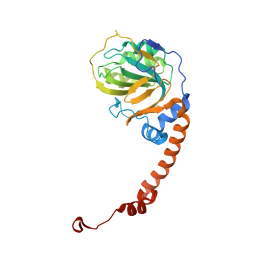
    4-Hydroxy-4-methyl-2-oxoglutarate/4-carboxy-4-hydroxy-2-oxoadipate (HMG/CHA) aldolase from Pseudomonas putida F1 catalyzes the last step of the bacterial protocatechuate 4,5-cleavage pathway. The preferred substrates of the enzyme are 2-keto-4-hydroxy acids with a 4-carboxylate substitution. The enzyme also exhibits oxaloacetate decarboxylation and pyruvate α-proton exchange activity. Sodium oxalate is a competitive inhibitor of the aldolase reaction. The pH dependence of k(cat)/K(m) and k(cat) for the enzyme is consistent with a single deprotonation with pK(a) values of 8.0 ± 0.1 and 7.0 ± 0.1 for free enzyme and enzyme substrate complex, respectively. The 1.8 Å x-ray structure shows a four-layered α-β-β-α sandwich structure with the active site at the interface of two adjacent subunits of a hexamer; this fold resembles the RNase E inhibitor, RraA, but is novel for an aldolase. The catalytic site contains a magnesium ion ligated by Asp-124 as well as three water molecules bound by Asp-102 and Glu-199'. A pyruvate molecule binds the magnesium ion through both carboxylate and keto oxygen atoms, completing the octahedral geometry. The carbonyl oxygen also forms hydrogen bonds with the guanadinium group of Arg-123, which site-directed mutagenesis confirms is essential for catalysis. A mechanism for HMG/CHA aldolase is proposed on the basis of the structure, kinetics, and previously established features of other aldolase mechanisms.


  • Organizational Affiliation
    • Department of Molecular and Cellular Biology, University of Guelph, Guelph, Ontario N1G 2W1, Canada.

Macromolecule Content 

  • Total Structure Weight: 25.98 kDa 
  • Atom Count: 1,938 
  • Modeled Residue Count: 235 
  • Deposited Residue Count: 238 
  • Unique protein chains: 1

Macromolecules

Find similar proteins by:|  3D Structure
Entity ID: 1
MoleculeChains  Sequence LengthOrganismDetailsImage
4-carboxy-4-hydroxy-2-oxoadipate aldolase/oxaloacetate decarboxylase238Pseudomonas putida F1Mutation(s): 0 
Gene Names: pput1361Pput_1361
EC: 4.1.3.17 (PDB Primary Data), 4.1.1.112 (UniProt)
UniProt
Find proteins for A5W059 (Pseudomonas putida (strain ATCC 700007 / DSM 6899 / JCM 31910 / BCRC 17059 / LMG 24140 / F1))
Explore A5W059 
Go to UniProtKB:  A5W059
Entity Groups
Sequence Clusters30% Identity50% Identity70% Identity90% Identity95% Identity100% Identity
UniProt GroupA5W059
Sequence Annotations
Expand
Reference Sequence

Experimental Data & Validation

Experimental Data

  • Method: X-RAY DIFFRACTION
  • Resolution: 1.82 Å
  • R-Value Free:  0.171 (Depositor), 0.177 (DCC) 
  • R-Value Work:  0.159 (Depositor), 0.166 (DCC) 
  • R-Value Observed: 0.159 (Depositor) 
Space Group: H 3 2
Unit Cell:
Length ( Å )Angle ( ˚ )
a = 111.18α = 90
b = 111.18β = 90
c = 139.273γ = 120
Software Package:
Software NamePurpose
PHASERphasing
REFMACrefinement
MxDCdata collection
HKL-2000data reduction
HKL-2000data scaling

Structure Validation

View Full Validation Report


Entry History 

Deposition Data

Revision History  (Full details and data files)

  • Version 1.0: 2010-09-15
    Type: Initial release
  • Version 1.1: 2011-07-13
    Changes: Source and taxonomy, Version format compliance
  • Version 1.2: 2023-09-06
    Changes: Data collection, Database references, Derived calculations, Refinement description
  • Version 2.0: 2023-11-15
    Changes: Atomic model, Data collection, Derived calculations