3L4V

Crystal complex of N-terminal Human Maltase-Glucoamylase with kotalanol


Experimental Data Snapshot

  • Method: X-RAY DIFFRACTION
  • Resolution: 2.10 Å
  • R-Value Free: 0.232 
  • R-Value Work: 0.182 
  • R-Value Observed: 0.185 

wwPDB Validation   3D Report Full Report


Ligand Structure Quality Assessment 


This is version 2.0 of the entry. See complete history


Literature

New glucosidase inhibitors from an ayurvedic herbal treatment for type 2 diabetes: structures and inhibition of human intestinal maltase-glucoamylase with compounds from Salacia reticulata.

Sim, L.Jayakanthan, K.Mohan, S.Nasi, R.Johnston, B.D.Pinto, B.M.Rose, D.R.

(2010) Biochemistry 49: 443-451

  • DOI: https://doi.org/10.1021/bi9016457
  • Primary Citation of Related Structures:  
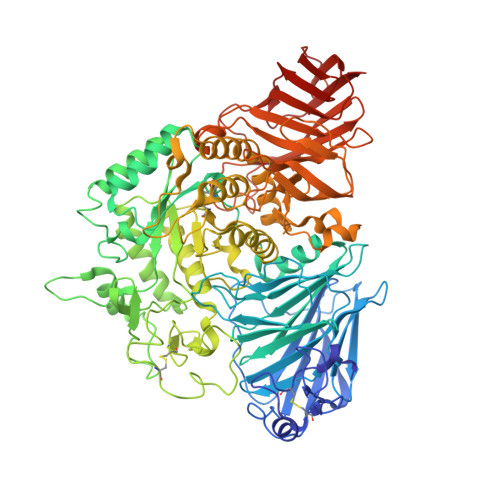
    3L4T, 3L4U, 3L4V, 3L4W, 3L4X, 3L4Y, 3L4Z

  • PubMed Abstract: 
  • An approach to controlling blood glucose levels in individuals with type 2 diabetes is to target alpha-amylases and intestinal glucosidases using alpha-glucosidase inhibitors acarbose and miglitol. One of the intestinal glucosidases targeted is the N-terminal catalytic domain of maltase-glucoamylase (ntMGAM), one of the four intestinal glycoside hydrolase 31 enzyme activities responsible for the hydrolysis of terminal starch products into glucose ...

    An approach to controlling blood glucose levels in individuals with type 2 diabetes is to target alpha-amylases and intestinal glucosidases using alpha-glucosidase inhibitors acarbose and miglitol. One of the intestinal glucosidases targeted is the N-terminal catalytic domain of maltase-glucoamylase (ntMGAM), one of the four intestinal glycoside hydrolase 31 enzyme activities responsible for the hydrolysis of terminal starch products into glucose. Here we present the X-ray crystallographic studies of ntMGAM in complex with a new class of alpha-glucosidase inhibitors derived from natural extracts of Salacia reticulata, a plant used traditionally in Ayuverdic medicine for the treatment of type 2 diabetes. Included in these extracts are the active compounds salacinol, kotalanol, and de-O-sulfonated kotalanol. This study reveals that de-O-sulfonated kotalanol is the most potent ntMGAM inhibitor reported to date (K(i) = 0.03 microM), some 2000-fold better than the compounds currently used in the clinic, and highlights the potential of the salacinol class of inhibitors as future drug candidates.


    Organizational Affiliation

    Ontario Cancer Institute and Department of Medical Biophysics, University of Toronto, 101 College Street, Toronto, ON, M5G 1L7 Canada.


Macromolecules
Find similar proteins by:  (by identity cutoff)  |  3D Structure
Entity ID: 1
MoleculeChainsSequence LengthOrganismDetailsImage
Maltase-glucoamylase, intestinalA875Homo sapiensMutation(s): 1 
Gene Names: MGAMGAMMGAML
EC: 3.2.1.20 (PDB Primary Data), 3.2.1.3 (PDB Primary Data)
UniProt & NIH Common Fund Data Resources
Find proteins for O43451 (Homo sapiens)
Explore O43451 
Go to UniProtKB:  O43451
PHAROS:  O43451
Entity Groups  
Sequence Clusters30% Identity50% Identity70% Identity90% Identity95% Identity100% Identity
UniProt GroupO43451
Protein Feature View
Expand
  • Reference Sequence
Oligosaccharides

Help

Entity ID: 2
MoleculeChainsChain Length2D DiagramGlycosylation3D Interactions
2-acetamido-2-deoxy-beta-D-glucopyranose-(1-4)-2-acetamido-2-deoxy-beta-D-glucopyranoseB, C 2N-Glycosylation Oligosaccharides Interaction
Glycosylation Resources
GlyTouCan:  G42666HT
GlyCosmos:  G42666HT
GlyGen:  G42666HT
Small Molecules
Ligands 2 Unique
IDChainsName / Formula / InChI Key2D Diagram3D Interactions
KTL
Query on KTL

Download Ideal Coordinates CCD File 
D [auth A](1S,2R,3R,4S)-1-{(1S)-2-[(2R,3S,4S)-3,4-dihydroxy-2-(hydroxymethyl)tetrahydrothiophenium-1-yl]-1-hydroxyethyl}-2,3,4,5-tetrahydroxypentyl sulfate
C12 H24 O12 S2
OMKXVFDVAGCPBS-QTIKZJLFSA-N
 Ligand Interaction
NAG
Query on NAG

Download Ideal Coordinates CCD File 
E [auth A]2-acetamido-2-deoxy-beta-D-glucopyranose
C8 H15 N O6
OVRNDRQMDRJTHS-FMDGEEDCSA-N
 Ligand Interaction
Binding Affinity Annotations 
IDSourceBinding Affinity
KTL PDBBind:  3L4V Ki: 190 (nM) from 1 assay(s)
Binding MOAD:  3L4V Ki: 190 (nM) from 1 assay(s)
Experimental Data & Validation

Experimental Data

  • Method: X-RAY DIFFRACTION
  • Resolution: 2.10 Å
  • R-Value Free: 0.232 
  • R-Value Work: 0.182 
  • R-Value Observed: 0.185 
  • Space Group: P 21 21 21
Unit Cell:
Length ( Å )Angle ( ˚ )
a = 91.278α = 90
b = 109.693β = 90
c = 110.075γ = 90
Software Package:
Software NamePurpose
DENZOdata reduction
SCALEPACKdata scaling
REFMACrefinement
PDB_EXTRACTdata extraction

Structure Validation

View Full Validation Report


Ligand Structure Quality Assessment 


Entry History 

Deposition Data

Revision History  (Full details and data files)

  • Version 1.0: 2010-02-09
    Type: Initial release
  • Version 1.1: 2011-07-13
    Changes: Version format compliance
  • Version 1.2: 2017-11-01
    Changes: Refinement description
  • Version 2.0: 2020-07-29
    Type: Remediation
    Reason: Carbohydrate remediation
    Changes: Advisory, Atomic model, Data collection, Database references, Derived calculations, Structure summary