3KDZ

X-ray crystal structure of a tyrosine aminomutase mutant construct with bound ligand


Experimental Data Snapshot

  • Method: X-RAY DIFFRACTION
  • Resolution: 2.20 Å
  • R-Value Free: 0.249 
  • R-Value Work: 0.205 
  • R-Value Observed: 0.205 

wwPDB Validation   3D Report Full Report


Ligand Structure Quality Assessment 


This is version 2.0 of the entry. See complete history


Literature

Probing the active site of MIO-dependent aminomutases, key catalysts in the biosynthesis of beta-amino acids incorporated in secondary metabolites

Cooke, H.A.Bruner, S.D.

(2010) Biopolymers 93: 802-810

  • DOI: https://doi.org/10.1002/bip.21500
  • Primary Citation of Related Structures:  
    3KDY, 3KDZ

  • PubMed Abstract: 

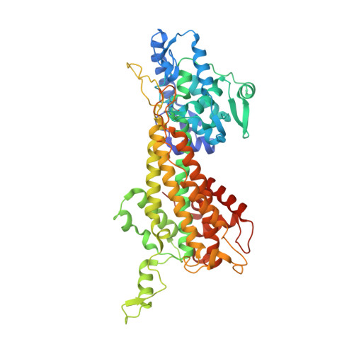
    The tyrosine aminomutase SgTAM produces (S)-ss-tyrosine from L-tyrosine in the biosynthesis of the enediyne antitumor antibiotic C-1027. This conversion is promoted by the methylideneimidazole-5-one (MIO) prosthetic group. MIO was first identified in the homologous family of ammonia lyases, which deaminate aromatic amino acids to form alpha,ss-unsaturated carboxylates. Studies of substrate specificity have been described for lyases but there have been limited reports in altering the substrate specificity of aminomutases. Furthermore, it remains unclear as to what structural properties are responsible for catalyzing the presumed readdition of the amino group into the alpha,ss-unsaturated intermediates to form ss-amino acids. Attempts to elucidate specificity and mechanistic determinants of SgTAM have also proved to be difficult as it is recalcitrant to perturbations to the active site via mutagenesis. An X-ray cocrystal structure of the SgTAM mutant of the catalytic base with L-tyrosine verified important substrate binding residues as well as the enzymatic base. Further mutagenesis revealed that removal of these crucial interactions renders the enzyme inactive. Proposed structural determinants for mutase activity probed via mutagenesis, time-point assays and X-ray crystallography revealed a complicated role for these residues in maintaining key quaternary structure properties that aid in catalysis.


  • Organizational Affiliation

    Department of Chemistry, Boston College, Chestnut Hill, MA 02467-3860, USA. bruner@ufl.edu


Macromolecules
Find similar proteins by:  (by identity cutoff)  |  3D Structure
Entity ID: 1
MoleculeChains Sequence LengthOrganismDetailsImage
Histidine ammonia-lyase
A, B
537Streptomyces globisporusMutation(s): 2 
Gene Names: sgcC4
EC: 4.3.1.3
UniProt
Find proteins for Q8GMG0 (Streptomyces globisporus)
Explore Q8GMG0 
Go to UniProtKB:  Q8GMG0
Entity Groups  
Sequence Clusters30% Identity50% Identity70% Identity90% Identity95% Identity100% Identity
UniProt GroupQ8GMG0
Sequence Annotations
Expand
  • Reference Sequence
Small Molecules
Modified Residues  1 Unique
IDChains TypeFormula2D DiagramParent
MDO
Query on MDO
A, B
L-PEPTIDE LINKINGC8 H11 N3 O3ALA, SER, GLY
Experimental Data & Validation

Experimental Data

  • Method: X-RAY DIFFRACTION
  • Resolution: 2.20 Å
  • R-Value Free: 0.249 
  • R-Value Work: 0.205 
  • R-Value Observed: 0.205 
  • Space Group: P 21 21 2
Unit Cell:
Length ( Å )Angle ( ˚ )
a = 92.722α = 90
b = 145.941β = 90
c = 74.812γ = 90
Software Package:
Software NamePurpose
CBASSdata collection
CNSrefinement
HKL-2000data reduction
SCALEPACKdata scaling
CNSphasing

Structure Validation

View Full Validation Report


Ligand Structure Quality Assessment 


Entry History 

Deposition Data

Revision History  (Full details and data files)

  • Version 1.0: 2010-07-07
    Type: Initial release
  • Version 1.1: 2011-07-13
    Changes: Version format compliance
  • Version 1.2: 2021-10-13
    Changes: Database references, Derived calculations
  • Version 1.3: 2023-09-06
    Changes: Data collection, Refinement description
  • Version 2.0: 2023-11-15
    Changes: Atomic model, Data collection, Derived calculations