3HHQ | pdb_00003hhq

Crystal structure of apo dUT1p from Saccharomyces cerevisiae


Experimental Data Snapshot

  • Method: X-RAY DIFFRACTION
  • Resolution: 2.00 Å
  • R-Value Free: 
    0.236 (Depositor), 0.234 (DCC) 
  • R-Value Work: 
    0.175 (Depositor), 0.173 (DCC) 
  • R-Value Observed: 
    0.178 (Depositor) 

Starting Model: experimental
View more details

wwPDB Validation 3D Report Full Report

Validation slider image for 3HHQ

This is version 1.3 of the entry. See complete history

Literature

Structure and activity of the Saccharomyces cerevisiae dUTP pyrophosphatase DUT1, an essential housekeeping enzyme.

Tchigvintsev, A.Singer, A.U.Flick, R.Petit, P.Brown, G.Evdokimova, E.Savchenko, A.Yakunin, A.F.

(2011) Biochem J 437: 243-253

  • DOI: https://doi.org/10.1042/BJ20110304
  • Primary Citation Related Structures: 
    3F4F, 3HHQ, 3P48

  • PubMed Abstract: 

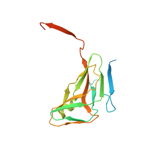
    Genomes of all free-living organisms encode the enzyme dUTPase (dUTP pyrophosphatase), which plays a key role in preventing uracil incorporation into DNA. In the present paper, we describe the biochemical and structural characterization of DUT1 (Saccharomyces cerevisiae dUTPase). The hydrolysis of dUTP by DUT1 was strictly dependent on a bivalent metal cation with significant activity observed in the presence of Mg2+, Co2+, Mn2+, Ni2+ or Zn2+. In addition, DUT1 showed a significant activity against another potentially mutagenic nucleotide: dITP. With both substrates, DUT1 demonstrated a sigmoidal saturation curve, suggesting a positive co-operativity between the subunits. The crystal structure of DUT1 was solved at 2 Å resolution (1 Å=0.1 nm) in an apo state and in complex with the non-hydrolysable substrate α,β-imido dUTP or dUMP product. Alanine-replacement mutagenesis of the active-site residues revealed seven residues important for activity including the conserved triad Asp87/Arg137/Asp85. The Y88A mutant protein was equally active against both dUTP and UTP, indicating that this conserved tyrosine residue is responsible for discrimination against ribonucleotides. The structure of DUT1 and site-directed mutagenesis support a role of the conserved Phe142 in the interaction with the uracil base. Our work provides further insight into the molecular mechanisms of substrate selectivity and catalysis of dUTPases.


  • Organizational Affiliation
    • Department of Chemical Engineering and Applied Chemistry, Banting and Best Department of Medical Research, Centre for Structural Proteomics in Toronto, University of Toronto, Toronto, Canada.

Macromolecule Content 

  • Total Structure Weight: 427.1 kDa 
  • Atom Count: 25,087 
  • Modeled Residue Count: 3,045 
  • Deposited Residue Count: 4,008 
  • Unique protein chains: 1

Macromolecules

Find similar proteins by:|  3D Structure
Entity ID: 1
MoleculeChains  Sequence LengthOrganismDetailsImage
Deoxyuridine 5'-triphosphate nucleotidohydrolase
A, B, C, D, E
A, B, C, D, E, F, G, H, I, J, K, L, M, N, O, P, Q, R, S, T, U, V, W, X
167Saccharomyces cerevisiaeMutation(s): 0 
Gene Names: DUT1dUT1pYBR1705YBR252W
EC: 3.6.1.23
UniProt
Find proteins for P33317 (Saccharomyces cerevisiae (strain ATCC 204508 / S288c))
Explore P33317 
Go to UniProtKB:  P33317
Entity Groups
Sequence Clusters30% Identity50% Identity70% Identity90% Identity95% Identity100% Identity
UniProt GroupP33317
Sequence Annotations
Expand
Reference Sequence

Small Molecules

Ligands 6 Unique
IDChains Name / Formula / InChI Key2D Diagram3D Interactions
PEG

Query on PEG


Download:Ideal Coordinates CCD File
AE [auth X]
BE [auth X]
GB [auth J]
HB [auth J]
NB [auth K]
AE [auth X],
BE [auth X],
GB [auth J],
HB [auth J],
NB [auth K],
YB [auth M],
ZA [auth H]
DI(HYDROXYETHYL)ETHER
C4 H10 O3
MTHSVFCYNBDYFN-UHFFFAOYSA-N
SO4

Query on SO4


Download:Ideal Coordinates CCD File
AB [auth I]
AC [auth N]
AD [auth S]
BA [auth B]
CA [auth B]
AB [auth I],
AC [auth N],
AD [auth S],
BA [auth B],
CA [auth B],
CB [auth J],
DD [auth T],
FC [auth O],
GA [auth D],
IB [auth K],
JB [auth K],
KD [auth U],
LA [auth E],
LC [auth P],
MC [auth P],
OA [auth F],
OD [auth V],
PA [auth F],
PC [auth Q],
PD [auth V],
QA [auth F],
QD [auth V],
RB [auth M],
SB [auth M],
UA [auth G],
VA [auth G],
VC [auth R],
WA [auth G],
XD [auth W],
Y [auth A],
YA [auth H],
YD [auth W],
Z [auth A],
ZB [auth N],
ZC [auth S]
SULFATE ION
O4 S
QAOWNCQODCNURD-UHFFFAOYSA-L
GOL

Query on GOL


Download:Ideal Coordinates CCD File
ND [auth U]GLYCEROL
C3 H8 O3
PEDCQBHIVMGVHV-UHFFFAOYSA-N
EDO

Query on EDO


Download:Ideal Coordinates CCD File
CD [auth S]
EB [auth J]
EC [auth N]
FB [auth J]
HC [auth O]
CD [auth S],
EB [auth J],
EC [auth N],
FB [auth J],
HC [auth O],
HD [auth T],
ID [auth T],
JA [auth D],
JC [auth O],
JD [auth T],
KA [auth D],
KB [auth K],
KC [auth O],
LB [auth K],
MB [auth K],
MD [auth U],
NA [auth E],
OC [auth P],
RC [auth Q],
SC [auth Q],
SD [auth V],
TA [auth F],
TC [auth Q],
TD [auth V],
UB [auth M],
UC [auth Q],
VB [auth M],
WB [auth M],
WD [auth V],
XA [auth G],
XB [auth M],
XC [auth R],
YC [auth R],
ZD [auth W]
1,2-ETHANEDIOL
C2 H6 O2
LYCAIKOWRPUZTN-UHFFFAOYSA-N
CL

Query on CL


Download:Ideal Coordinates CCD File
BB [auth I]
CC [auth N]
DA [auth B]
DB [auth J]
DC [auth N]
BB [auth I],
CC [auth N],
DA [auth B],
DB [auth J],
DC [auth N],
EA [auth C],
GD [auth T],
HA [auth D],
IA [auth D],
LD [auth U],
MA [auth E],
OB [auth L],
QB [auth L],
QC [auth Q],
RA [auth F],
VD [auth V],
WC [auth R]
CHLORIDE ION
Cl
VEXZGXHMUGYJMC-UHFFFAOYSA-M
NA

Query on NA


Download:Ideal Coordinates CCD File
AA [auth A]
BC [auth N]
BD [auth S]
ED [auth T]
FA [auth C]
AA [auth A],
BC [auth N],
BD [auth S],
ED [auth T],
FA [auth C],
FD [auth T],
GC [auth O],
IC [auth O],
NC [auth P],
PB [auth L],
RD [auth V],
SA [auth F],
TB [auth M],
UD [auth V]
SODIUM ION
Na
FKNQFGJONOIPTF-UHFFFAOYSA-N

Experimental Data & Validation

Experimental Data

  • Method: X-RAY DIFFRACTION
  • Resolution: 2.00 Å
  • R-Value Free:  0.236 (Depositor), 0.234 (DCC) 
  • R-Value Work:  0.175 (Depositor), 0.173 (DCC) 
  • R-Value Observed: 0.178 (Depositor) 
Space Group: P 1
Unit Cell:
Length ( Å )Angle ( ˚ )
a = 95.461α = 98.79
b = 95.466β = 97.42
c = 97.594γ = 107.13
Software Package:
Software NamePurpose
CrystalCleardata collection
PHASERphasing
MOLREPphasing
REFMACrefinement
HKL-2000data reduction
HKL-2000data scaling

Structure Validation

View Full Validation Report


Entry History 

Deposition Data

Revision History  (Full details and data files)

  • Version 1.0: 2009-06-16
    Type: Initial release
  • Version 1.1: 2011-07-13
    Changes: Advisory, Refinement description, Version format compliance
  • Version 1.2: 2012-10-10
    Changes: Database references
  • Version 1.3: 2023-09-06
    Changes: Data collection, Database references, Derived calculations, Refinement description