3H6G

Crystal structure of the GluR6 amino terminal domain dimer assembly


Experimental Data Snapshot

  • Method: X-RAY DIFFRACTION
  • Resolution: 2.70 Å
  • R-Value Free: 0.227 
  • R-Value Work: 0.204 
  • R-Value Observed: 0.205 

wwPDB Validation   3D Report Full Report


Ligand Structure Quality Assessment 


This is version 1.4 of the entry. See complete history


Literature

The N-terminal domain of GluR6-subtype glutamate receptor ion channels.

Kumar, J.Schuck, P.Jin, R.Mayer, M.L.

(2009) Nat Struct Mol Biol 16: 631-638

  • DOI: https://doi.org/10.1038/nsmb.1613
  • Primary Citation of Related Structures:  
    3H6G, 3H6H

  • PubMed Abstract: 

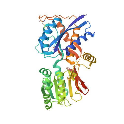
    The amino-terminal domain (ATD) of glutamate receptor ion channels, which controls their selective assembly into AMPA, kainate and NMDA receptor subtypes, is also the site of action of NMDA receptor allosteric modulators. Here we report the crystal structure of the ATD from the kainate receptor GluR6. The ATD forms dimers in solution at micromolar protein concentrations and crystallizes as a dimer. Unexpectedly, each subunit adopts an intermediate extent of domain closure compared to the apo and ligand-bound complexes of LIVBP and G protein-coupled glutamate receptors (mGluRs), and the dimer assembly has a markedly different conformation from that found in mGluRs. This conformation is stabilized by contacts between large hydrophobic patches in the R2 domain that are absent in NMDA receptors, suggesting that the ATDs of individual glutamate receptor ion channels have evolved into functionally distinct families.


  • Organizational Affiliation

    Laboratory of Cellular and Molecular Neurophysiology, Porter Neuroscience Research Center, National Institute of Child Health and Human Development, Bethesda, Maryland, USA.


Macromolecules
Find similar proteins by:  (by identity cutoff)  |  3D Structure
Entity ID: 1
MoleculeChains Sequence LengthOrganismDetailsImage
Glutamate receptor, ionotropic kainate 2
A, B
395Rattus norvegicusMutation(s): 0 
Gene Names: Glur6Grik2
UniProt
Find proteins for P42260 (Rattus norvegicus)
Explore P42260 
Go to UniProtKB:  P42260
Entity Groups  
Sequence Clusters30% Identity50% Identity70% Identity90% Identity95% Identity100% Identity
UniProt GroupP42260
Sequence Annotations
Expand
  • Reference Sequence
Small Molecules
Ligands 3 Unique
IDChains Name / Formula / InChI Key2D Diagram3D Interactions
NAG
Query on NAG

Download Ideal Coordinates CCD File 
C [auth A]
D [auth A]
E [auth A]
F [auth A]
G [auth A]
C [auth A],
D [auth A],
E [auth A],
F [auth A],
G [auth A],
J [auth B],
K [auth B],
L [auth B]
2-acetamido-2-deoxy-beta-D-glucopyranose
C8 H15 N O6
OVRNDRQMDRJTHS-FMDGEEDCSA-N
TLA
Query on TLA

Download Ideal Coordinates CCD File 
H [auth A],
M [auth B]
L(+)-TARTARIC ACID
C4 H6 O6
FEWJPZIEWOKRBE-JCYAYHJZSA-N
CA
Query on CA

Download Ideal Coordinates CCD File 
I [auth A],
N [auth B]
CALCIUM ION
Ca
BHPQYMZQTOCNFJ-UHFFFAOYSA-N
Experimental Data & Validation

Experimental Data

  • Method: X-RAY DIFFRACTION
  • Resolution: 2.70 Å
  • R-Value Free: 0.227 
  • R-Value Work: 0.204 
  • R-Value Observed: 0.205 
  • Space Group: P 61
Unit Cell:
Length ( Å )Angle ( ˚ )
a = 172.074α = 90
b = 172.074β = 90
c = 111.547γ = 120
Software Package:
Software NamePurpose
HKL-2000data collection
PHASERphasing
PHENIXrefinement
HKL-2000data reduction
HKL-2000data scaling

Structure Validation

View Full Validation Report


Ligand Structure Quality Assessment 


Entry History 

Deposition Data

Revision History  (Full details and data files)

  • Version 1.0: 2009-05-26
    Type: Initial release
  • Version 1.1: 2011-07-13
    Changes: Non-polymer description, Version format compliance
  • Version 1.2: 2020-07-29
    Type: Remediation
    Reason: Carbohydrate remediation
    Changes: Advisory, Data collection, Database references, Derived calculations, Structure summary
  • Version 1.3: 2021-03-31
    Changes: Source and taxonomy, Structure summary
  • Version 1.4: 2023-09-06
    Changes: Data collection, Database references, Refinement description