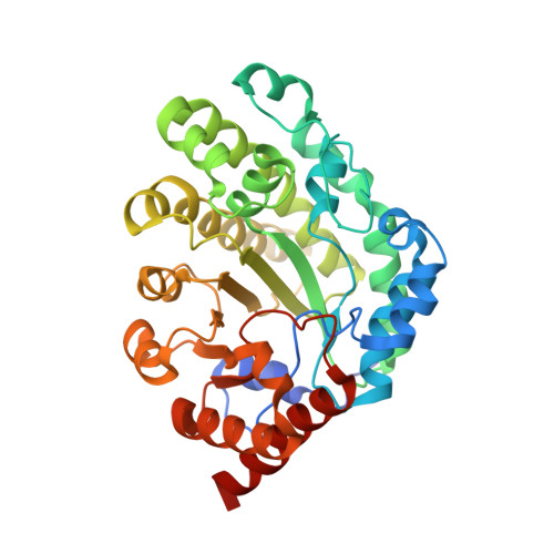
Substrate shuttling between active sites of uroporphyrinogen decarboxylase is not required to generate coproporphyrinogen.
Phillips, J.D., Warby, C.A., Whitby, F.G., Kushner, J.P., Hill, C.P.(2009) J Mol Biology 389: 306-314
- PubMed: 19362562
- DOI: https://doi.org/10.1016/j.jmb.2009.04.013
- Primary Citation of Related Structures:
3GVQ, 3GVR, 3GVV, 3GVW - PubMed Abstract:
Uroporphyrinogen decarboxylase (URO-D; EC 4.1.1.37), the fifth enzyme of the heme biosynthetic pathway, is required for the production of heme, vitamin B12, siroheme, and chlorophyll precursors. URO-D catalyzes the sequential decarboxylation of four acetate side chains in the pyrrole groups of uroporphyrinogen to produce coproporphyrinogen. URO-D is a stable homodimer, with the active-site clefts of the two subunits adjacent to each other. It has been hypothesized that the two catalytic centers interact functionally, perhaps by shuttling of reaction intermediates between subunits. We tested this hypothesis by construction of a single-chain protein (single-chain URO-D) in which the two subunits were connected by a flexible linker. The crystal structure of this protein was shown to be superimposable with wild-type activity and to have comparable catalytic activity. Mutations that impaired one or the other of the two active sites of single-chain URO-D resulted in approximately half of wild-type activity. The distributions of reaction intermediates were the same for mutant and wild-type sequences and were unaltered in a competition experiment using I and III isomer substrates. These observations indicate that communication between active sites is not required for enzyme function and suggest that the dimeric structure of URO-D is required to achieve conformational stability and to create a large active-site cleft.
- Department of Medicine, University of Utah School of Medicine, 5C330 SOM, 30 North 1900 East, Salt Lake City, UT 84132, USA. john.phillips@hsc.utah.edu
Organizational Affiliation:


