Kinetic, structural, and EPR studies reveal that aldehyde oxidoreductase from Desulfovibrio gigas does not need a sulfido ligand for catalysis and give evidence for a direct Mo-C interaction in a biological system.
Santos-Silva, T., Ferroni, F., Thapper, A., Marangon, J., Gonzalez, P.J., Rizzi, A.C., Moura, I., Moura, J.J., Romao, M.J., Brondino, C.D.(2009) J Am Chem Soc 131: 7990-7998
- PubMed: 19459677 Search on PubMed
- DOI: https://doi.org/10.1021/ja809448r
- Primary Citation Related Structures:
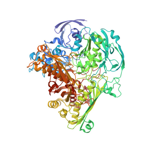
3FAH, 3FC4 - PubMed Abstract:
Aldehyde oxidoreductase from Desulfovibrio gigas (DgAOR) is a member of the xanthine oxidase (XO) family of mononuclear Mo-enzymes that catalyzes the oxidation of aldehydes to carboxylic acids. The molybdenum site in the enzymes of the XO family shows a distorted square pyramidal geometry in which two ligands, a hydroxyl/water molecule (the catalytic labile site) and a sulfido ligand, have been shown to be essential for catalysis. We report here steady-state kinetic studies of DgAOR with the inhibitors cyanide, ethylene glycol, glycerol, and arsenite, together with crystallographic and EPR studies of the enzyme after reaction with the two alcohols. In contrast to what has been observed in other members of the XO family, cyanide, ethylene glycol, and glycerol are reversible inhibitors of DgAOR. Kinetic data with both cyanide and samples prepared from single crystals confirm that DgAOR does not need a sulfido ligand for catalysis and confirm the absence of this ligand in the coordination sphere of the molybdenum atom in the active enzyme. Addition of ethylene glycol and glycerol to dithionite-reduced DgAOR yields rhombic Mo(V) EPR signals, suggesting that the nearly square pyramidal coordination of the active enzyme is distorted upon alcohol inhibition. This is in agreement with the X-ray structure of the ethylene glycol and glycerol-inhibited enzyme, where the catalytically labile OH/OH(2) ligand is lost and both alcohols coordinate the Mo site in a eta(2) fashion. The two adducts present a direct interaction between the molybdenum and one of the carbon atoms of the alcohol moiety, which constitutes the first structural evidence for such a bond in a biological system.
- REQUIMTE, Departamento de Química, Centro de Química Fina e Biotecnologia, Faculdade de Ciências e Tecnologia, Universidade Nova de Lisboa, 2829-516 Caparica, Portugal.
Organizational Affiliation: