3F5X

CDK-2-Cyclin complex with indazole inhibitor 9 bound at its active site


Experimental Data Snapshot

  • Method: X-RAY DIFFRACTION
  • Resolution: 2.40 Å
  • R-Value Free: 0.283 
  • R-Value Work: 0.266 
  • R-Value Observed: 0.266 

wwPDB Validation   3D Report Full Report


Ligand Structure Quality Assessment 


This is version 1.1 of the entry. See complete history


Literature

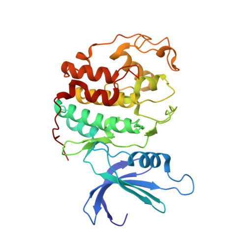
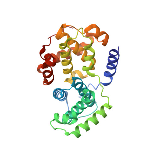
2-(6-Phenyl-1H-indazol-3-yl)-1H-benzo[d]imidazoles: Design and synthesis of a potent and isoform selective PKC-zeta inhibitor.

Trujillo, J.I.Kiefer, J.R.Huang, W.Thorarensen, A.Xing, L.Caspers, N.L.Day, J.E.Mathis, K.J.Kretzmer, K.K.Reitz, B.A.Weinberg, R.A.Stegeman, R.A.Wrightstone, A.Christine, L.Compton, R.Li, X.

(2009) Bioorg Med Chem Lett 19: 908-911

  • DOI: 10.1016/j.bmcl.2008.11.105
  • Primary Citation of Related Structures:  
    3EZR, 3EZV, 3F5X

  • PubMed Abstract: 
  • The inhibition of PKC-zeta has been proposed to be a potential drug target for immune and inflammatory diseases. A series of 2-(6-phenyl-1H indazol-3-yl)-1H-benzo[d]imidazoles with initial high crossover to CDK-2 has been optimized to afford potent and selective inhibitors of protein kinase c-zeta (PKC-zeta) ...

    The inhibition of PKC-zeta has been proposed to be a potential drug target for immune and inflammatory diseases. A series of 2-(6-phenyl-1H indazol-3-yl)-1H-benzo[d]imidazoles with initial high crossover to CDK-2 has been optimized to afford potent and selective inhibitors of protein kinase c-zeta (PKC-zeta). The determination of the crystal structures of key inhibitor:CDK-2 complexes informed the design and analysis of the series. The most selective and potent analog was identified by variation of the aryl substituent at the 6-position of the indazole template to give a 4-NH(2) derivative. The analog displays good selectivity over other PKC isoforms (alpha, betaII, gamma, delta, epsilon, mu, theta, eta and iota/lambda) and CDK-2, however it displays marginal selectivity against a panel of other kinases (37 profiled).


    Organizational Affiliation

    Department of Medicinal Chemistry, Pfizer Global Research and Development, 700 Chesterfield Pkwy, AA236, Chesterfield, MO 63017, USA. john.i.trujillo@pfizer.com


Macromolecules
Find similar proteins by:  (by identity cutoff)  |  3D Structure
Entity ID: 1
MoleculeChainsSequence LengthOrganismDetailsImage
Cell division protein kinase 2A, C298Homo sapiensMutation(s): 0 
Gene Names: CDK2CDKN2
EC: 2.7.11.22
UniProt & NIH Common Fund Data Resources
Find proteins for P24941 (Homo sapiens)
Explore P24941 
Go to UniProtKB:  P24941
PHAROS:  P24941
Entity Groups  
Sequence Clusters30% Identity50% Identity70% Identity90% Identity95% Identity100% Identity
UniProt GroupP24941
Protein Feature View
Expand
  • Reference Sequence
Find similar proteins by:  (by identity cutoff)  |  3D Structure
Entity ID: 2
MoleculeChainsSequence LengthOrganismDetailsImage
Cyclin-A2B, D256Homo sapiensMutation(s): 0 
Gene Names: CCNA2CCN1CCNA
UniProt & NIH Common Fund Data Resources
Find proteins for P20248 (Homo sapiens)
Explore P20248 
Go to UniProtKB:  P20248
PHAROS:  P20248
Entity Groups  
Sequence Clusters30% Identity50% Identity70% Identity90% Identity95% Identity100% Identity
UniProt GroupP20248
Protein Feature View
Expand
  • Reference Sequence
Small Molecules
Ligands 3 Unique
IDChainsName / Formula / InChI Key2D Diagram3D Interactions
EZV
Query on EZV

Download Ideal Coordinates CCD File 
F [auth A],
H [auth C]
4-{3-[7-(4-methylpiperazin-1-yl)-1H-benzimidazol-2-yl]-1H-indazol-6-yl}aniline
C25 H25 N7
LPSCCOBGZSKECZ-UHFFFAOYSA-N
 Ligand Interaction
SO4
Query on SO4

Download Ideal Coordinates CCD File 
I [auth D]SULFATE ION
O4 S
QAOWNCQODCNURD-UHFFFAOYSA-L
 Ligand Interaction
GOL
Query on GOL

Download Ideal Coordinates CCD File 
E [auth A],
G [auth C]
GLYCEROL
C3 H8 O3
PEDCQBHIVMGVHV-UHFFFAOYSA-N
 Ligand Interaction
Binding Affinity Annotations 
IDSourceBinding Affinity
EZV Binding MOAD:  3F5X IC50: 1040 (nM) from 1 assay(s)
Experimental Data & Validation

Experimental Data

  • Method: X-RAY DIFFRACTION
  • Resolution: 2.40 Å
  • R-Value Free: 0.283 
  • R-Value Work: 0.266 
  • R-Value Observed: 0.266 
  • Space Group: P 62 2 2
Unit Cell:
Length ( Å )Angle ( ˚ )
a = 184.051α = 90
b = 184.051β = 90
c = 214.848γ = 120
Software Package:
Software NamePurpose
REFMACrefinement
PDB_EXTRACTdata extraction

Structure Validation

View Full Validation Report


Ligand Structure Quality Assessment 


Entry History 

Revision History  (Full details and data files)

  • Version 1.0: 2009-02-03
    Type: Initial release
  • Version 1.1: 2011-07-13
    Changes: Non-polymer description, Version format compliance