3EZV | pdb_00003ezv

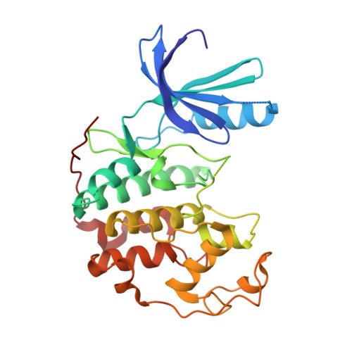
CDK-2 with indazole inhibitor 9 bound at its active site


Experimental Data Snapshot

  • Method: X-RAY DIFFRACTION
  • Resolution: 1.99 Å
  • R-Value Free: 
    0.244 (Depositor), 0.300 (DCC) 
  • R-Value Work: 
    0.227 (Depositor), 0.304 (DCC) 
  • R-Value Observed: 
    0.228 (Depositor) 

wwPDB Validation 3D Report Full Report

Validation slider image for 3EZV

Ligand Structure Quality Assessment 


This is version 1.2 of the entry. See complete history. 

Literature

2-(6-Phenyl-1H-indazol-3-yl)-1H-benzo[d]imidazoles: Design and synthesis of a potent and isoform selective PKC-zeta inhibitor

Trujillo, J.I., Kiefer, J.R., Huang, W., Thorarensen, A., Xing, L., Caspers, N.L., Day, J.E., Mathis, K.J., Kretzmer, K.K., Reitz, B.A., Weinberg, R.A., Stegeman, R.A., Wrightstone, A., Christine, L., Compton, R., Li, X.

(2009) Bioorg Med Chem Lett 19: 908-911

  • DOI: https://doi.org/10.1016/j.bmcl.2008.11.105
  • Primary Citation Related Structures: 
    3EZR, 3EZV, 3F5X

  • PubMed Abstract: 

    The inhibition of PKC-zeta has been proposed to be a potential drug target for immune and inflammatory diseases. A series of 2-(6-phenyl-1H indazol-3-yl)-1H-benzo[d]imidazoles with initial high crossover to CDK-2 has been optimized to afford potent and selective inhibitors of protein kinase c-zeta (PKC-zeta). The determination of the crystal structures of key inhibitor:CDK-2 complexes informed the design and analysis of the series. The most selective and potent analog was identified by variation of the aryl substituent at the 6-position of the indazole template to give a 4-NH(2) derivative. The analog displays good selectivity over other PKC isoforms (alpha, betaII, gamma, delta, epsilon, mu, theta, eta and iota/lambda) and CDK-2, however it displays marginal selectivity against a panel of other kinases (37 profiled).


  • Organizational Affiliation: 
    • Department of Medicinal Chemistry, Pfizer Global Research and Development, 700 Chesterfield Pkwy, AA236, Chesterfield, MO 63017, USA. john.i.trujillo@pfizer.com

Macromolecule Content 

  • Total Structure Weight: 34.54 kDa 
  • Atom Count: 2,402 
  • Modeled Residue Count: 277 
  • Deposited Residue Count: 300 
  • Unique protein chains: 1

Macromolecules

Find similar proteins by:|  3D Structure
Entity ID: 1
MoleculeChains  Sequence LengthOrganismDetailsImage
Cell division protein kinase 2300Homo sapiensMutation(s): 0 
Gene Names: CDK2
EC: 2.7.11.22
UniProt & NIH Common Fund Data Resources
Find proteins for P24941 (Homo sapiens)
Explore P24941 
Go to UniProtKB:  P24941
PHAROS:  P24941
GTEx:  ENSG00000123374 
Entity Groups
Sequence Clusters30% Identity50% Identity70% Identity90% Identity95% Identity100% Identity
UniProt GroupP24941
Sequence Annotations
Expand
Reference Sequence

Small Molecules

Ligands 1 Unique
IDChains Name / Formula / InChI Key2D Diagram3D Interactions
EZV

Query on EZV


Download:Ideal Coordinates CCD File
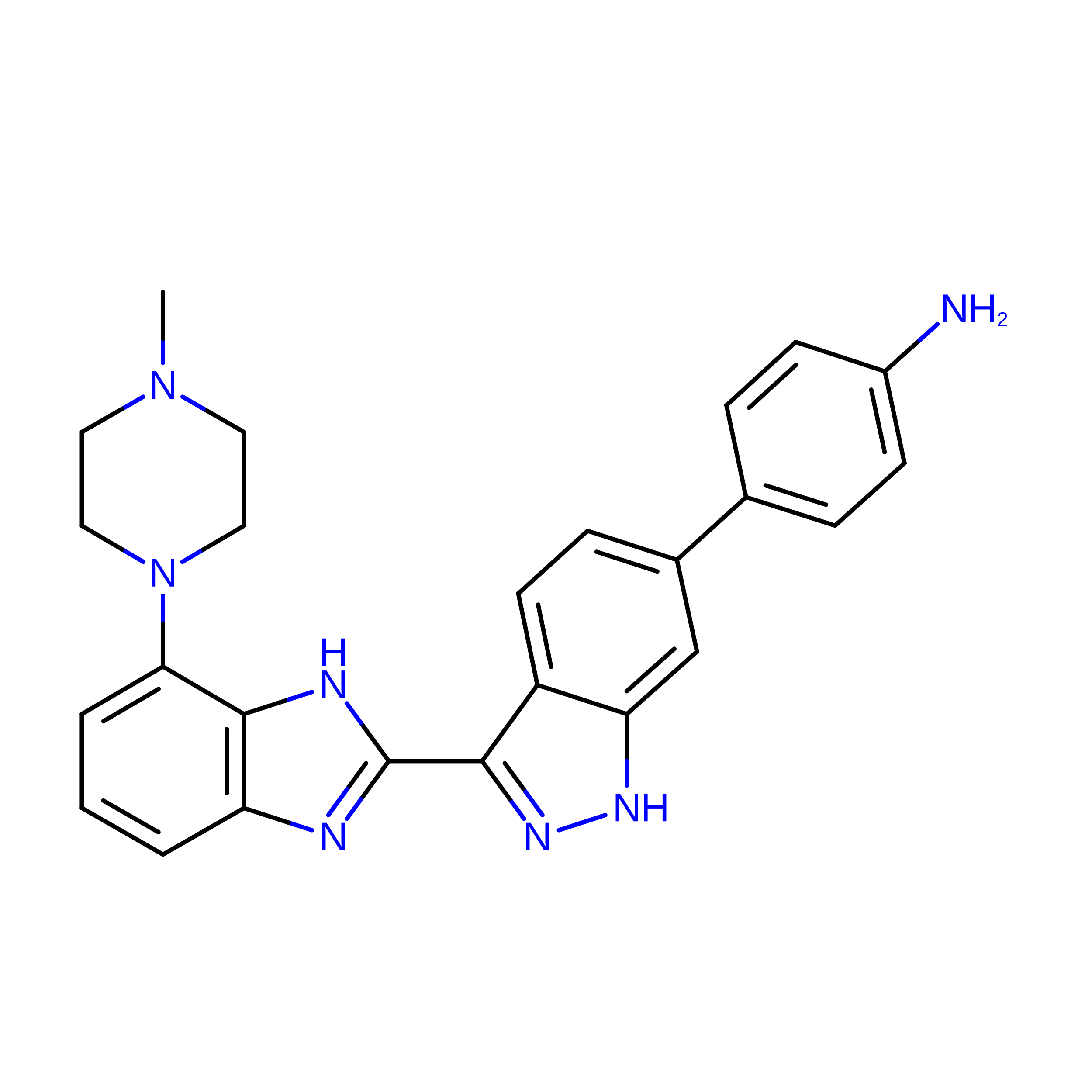
B [auth A]4-{3-[7-(4-methylpiperazin-1-yl)-1H-benzimidazol-2-yl]-1H-indazol-6-yl}aniline
C25 H25 N7
LPSCCOBGZSKECZ-UHFFFAOYSA-N

Experimental Data & Validation

Experimental Data

  • Method: X-RAY DIFFRACTION
  • Resolution: 1.99 Å
  • R-Value Free:  0.244 (Depositor), 0.300 (DCC) 
  • R-Value Work:  0.227 (Depositor), 0.304 (DCC) 
  • R-Value Observed: 0.228 (Depositor) 
Space Group: P 21 21 21
Unit Cell:
Length ( Å )Angle ( ˚ )
a = 53.602α = 90
b = 71.747β = 90
c = 71.922γ = 90
Software Package:
Software NamePurpose
REFMACrefinement
PDB_EXTRACTdata extraction

Structure Validation

View Full Validation Report


Ligand Structure Quality Assessment 


Entry History 

Revision History  (Full details and data files)

  • Version 1.0: 2009-02-03
    Type: Initial release
  • Version 1.1: 2011-07-13
    Changes: Version format compliance
  • Version 1.2: 2023-12-27
    Changes: Data collection, Database references, Derived calculations