3EVJ

Intermediate structure of antithrombin bound to the natural pentasaccharide


Experimental Data Snapshot

  • Method: X-RAY DIFFRACTION
  • Resolution: 3.00 Å
  • R-Value Free: 0.289 
  • R-Value Work: 0.231 
  • R-Value Observed: 0.231 

wwPDB Validation   3D Report Full Report


Ligand Structure Quality Assessment 


This is version 2.1 of the entry. See complete history


Literature

The critical role of hinge-region expulsion in the induced-fit heparin binding mechanism of antithrombin.

Langdown, J.Belzar, K.J.Savory, W.J.Baglin, T.P.Huntington, J.A.

(2009) J Mol Biol 386: 1278-1289

  • DOI: https://doi.org/10.1016/j.jmb.2009.01.028
  • Primary Citation of Related Structures:  
    3EVJ

  • PubMed Abstract: 

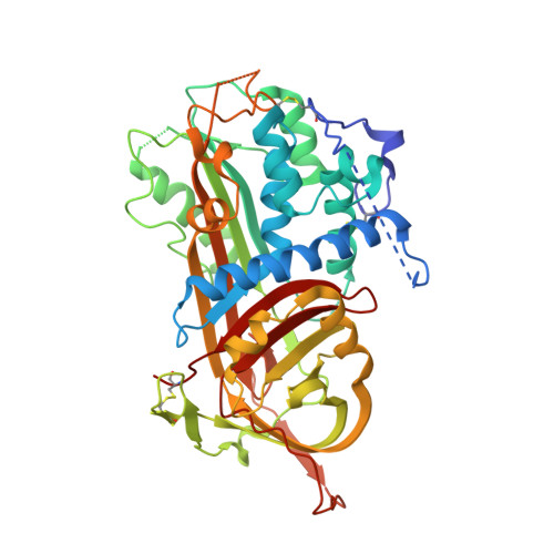
    Antithrombin (AT) is the most important inhibitor of coagulation proteases. Its activity is stimulated by glycosaminoglycans, such as heparin, through allosteric and template mechanisms. AT utilises an induced-fit mechanism to bind with high affinity to a pentasaccharide sequence found in about one-third of heparin chains. The conformational changes behind this mechanism have been characterised by several crystal structures of AT in the absence and in the presence of pentasaccharide. Pentasaccharide binding ultimately results in a conformational change that improves affinity by about 1000-fold. Crystal structures show several differences, including the expulsion of the hinge region of the reactive centre loop from beta-sheet A, which is known to be critical for the allosteric activation of AT. Here, we present data that reveal an energetically distinct intermediate on the path to full activation where the majority of conformational changes have already occurred. A crystal structure of this intermediate shows that the hinge region is in a native-like state in spite of having the pentasaccharide bound in the normal fashion. We engineered a disulfide bond to lock the hinge in its native position to determine the energetic contributions of the initial and final conformational events. Approximately 60% of the free-energy contribution of conformational change is provided by the final step of hinge-region expulsion and subsequent closure of the main beta-sheet A. A new analysis of the individual structural changes provides a plausible mechanism for propagation of conformational change from the heparin binding site to the remote hinge region in beta-sheet A.


  • Organizational Affiliation

    Department of Haematology, Cambridge Institute for Medical Research, University of Cambridge, Wellcome Trust/MRC Building, Hills Road, Cambridge CB2 0XY, UK.


Macromolecules
Find similar proteins by:  (by identity cutoff)  |  3D Structure
Entity ID: 1
MoleculeChains Sequence LengthOrganismDetailsImage
Antithrombin-IIIA [auth I],
B [auth L]
432Homo sapiensMutation(s): 0 
UniProt & NIH Common Fund Data Resources
Find proteins for P01008 (Homo sapiens)
Explore P01008 
Go to UniProtKB:  P01008
PHAROS:  P01008
GTEx:  ENSG00000117601 
Entity Groups  
Sequence Clusters30% Identity50% Identity70% Identity90% Identity95% Identity100% Identity
UniProt GroupP01008
Sequence Annotations
Expand
  • Reference Sequence
Oligosaccharides

Help

Entity ID: 2
MoleculeChains Length2D Diagram Glycosylation3D Interactions
alpha-D-mannopyranose-(1-4)-2-acetamido-2-deoxy-beta-D-glucopyranose-(1-4)-2-acetamido-2-deoxy-beta-D-glucopyranoseC [auth A]3N-Glycosylation
Glycosylation Resources
GlyTouCan:  G62182OO
GlyCosmos:  G62182OO
GlyGen:  G62182OO
Entity ID: 3
MoleculeChains Length2D Diagram Glycosylation3D Interactions
2-acetamido-2-deoxy-beta-D-glucopyranose-(1-4)-2-acetamido-2-deoxy-beta-D-glucopyranoseD [auth B],
E [auth C]
2N-Glycosylation
Glycosylation Resources
GlyTouCan:  G42666HT
GlyCosmos:  G42666HT
GlyGen:  G42666HT
Entity ID: 4
MoleculeChains Length2D Diagram Glycosylation3D Interactions
alpha-D-mannopyranose-(1-3)-[alpha-D-mannopyranose-(1-6)]alpha-D-mannopyranose-(1-4)-2-acetamido-2-deoxy-beta-D-glucopyranose-(1-4)-2-acetamido-2-deoxy-beta-D-glucopyranoseF [auth D]5N-Glycosylation
Glycosylation Resources
GlyTouCan:  G21381MC
GlyCosmos:  G21381MC
GlyGen:  G21381MC
Entity ID: 5
MoleculeChains Length2D Diagram Glycosylation3D Interactions
2-deoxy-6-O-sulfo-2-(sulfoamino)-alpha-D-glucopyranose-(1-4)-beta-D-glucopyranuronic acid-(1-4)-2-deoxy-3,6-di-O-sulfo-2-(sulfoamino)-alpha-D-glucopyranose-(1-4)-2-O-sulfo-alpha-L-idopyranuronic acid-(1-4)-methyl 2-deoxy-6-O-sulfo-2-(sulfoamino)-alpha-D-glucopyranosideG [auth E],
H [auth F]
5N/A
Glycosylation Resources
GlyTouCan:  G25282IW
GlyCosmos:  G25282IW
Small Molecules
Ligands 1 Unique
IDChains Name / Formula / InChI Key2D Diagram3D Interactions
NAG
Query on NAG

Download Ideal Coordinates CCD File 
I,
J [auth L]
2-acetamido-2-deoxy-beta-D-glucopyranose
C8 H15 N O6
OVRNDRQMDRJTHS-FMDGEEDCSA-N
Biologically Interesting Molecules (External Reference) 1 Unique
Experimental Data & Validation

Experimental Data

  • Method: X-RAY DIFFRACTION
  • Resolution: 3.00 Å
  • R-Value Free: 0.289 
  • R-Value Work: 0.231 
  • R-Value Observed: 0.231 
  • Space Group: P 1 21 1
Unit Cell:
Length ( Å )Angle ( ˚ )
a = 65.846α = 90
b = 87.055β = 106.18
c = 92.383γ = 90
Software Package:
Software NamePurpose
ADSCdata collection
MOLREPphasing
CNSrefinement
MOSFLMdata reduction
SCALAdata scaling

Structure Validation

View Full Validation Report


Ligand Structure Quality Assessment 


Entry History 

Deposition Data

Revision History  (Full details and data files)

  • Version 1.0: 2008-10-21
    Type: Initial release
  • Version 1.1: 2011-07-13
    Changes: Non-polymer description, Version format compliance
  • Version 1.2: 2017-06-21
    Changes: Database references
  • Version 1.3: 2017-07-12
    Changes: Advisory, Structure summary
  • Version 2.0: 2020-07-29
    Type: Remediation
    Reason: Carbohydrate remediation
    Changes: Advisory, Atomic model, Data collection, Derived calculations, Non-polymer description, Structure summary
  • Version 2.1: 2023-09-06
    Changes: Advisory, Data collection, Database references, Refinement description, Structure summary