3EPI | pdb_00003epi

Structure of Human DNA Polymerase Iota complexed with N2-ethylguanine and incoming TTP


Experimental Data Snapshot

  • Method: X-RAY DIFFRACTION
  • Resolution: 2.90 Å
  • R-Value Free: 
    0.282 (Depositor), 0.236 (DCC) 
  • R-Value Work: 
    0.236 (Depositor), 0.250 (DCC) 
  • R-Value Observed: 
    0.239 (Depositor) 

Starting Model: experimental
View more details

wwPDB Validation 3D Report Full Report

Validation slider image for 3EPI

This is version 1.2 of the entry. See complete history

Literature

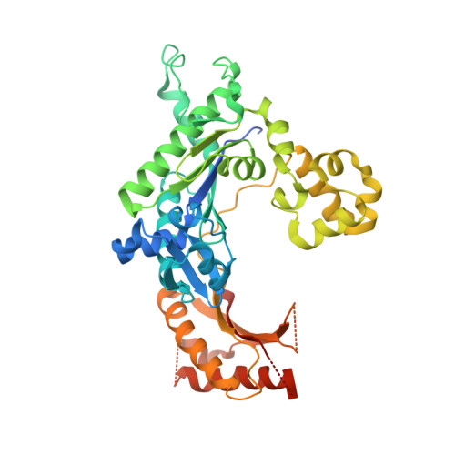
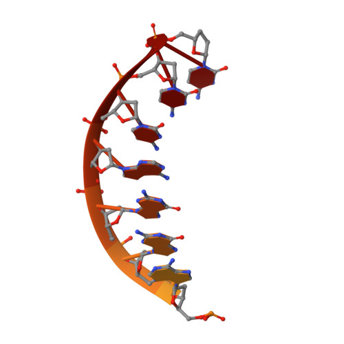
Lesion bypass of N2-ethylguanine by human DNA polymerase iota.

Pence, M.G.Blans, P.Zink, C.N.Hollis, T.Fishbein, J.C.Perrino, F.W.

(2009) J Biological Chem 284: 1732-1740

  • DOI: https://doi.org/10.1074/jbc.M807296200
  • Primary Citation Related Structures: 
    3EPG, 3EPI

  • PubMed Abstract: 

    Nucleotide incorporation and extension opposite N2-ethyl-Gua by DNA polymerase iota was measured and structures of the DNA polymerase iota-N2-ethyl-Gua complex with incoming nucleotides were solved. Efficiency and fidelity of DNA polymerase iota opposite N2-ethyl-Gua was determined by steady state kinetic analysis with Mg2+ or Mn2+ as the activating metal. DNA polymerase iota incorporates dCMP opposite N2-ethyl-Gua and unadducted Gua with similar efficiencies in the presence of Mg2+ and with greater efficiencies in the presence of Mn2+. However, the fidelity of nucleotide incorporation by DNA polymerase iota opposite N2-ethyl-Gua and Gua using Mn2+ is lower relative to that using Mg2+ indicating a metal-dependent effect. DNA polymerase iota extends from the N2-ethyl-Gua:Cyt 3' terminus more efficiently than from the Gua:Cyt base pair. Together these kinetic data indicate that the DNA polymerase iota catalyzed reaction is well suited for N(2)-ethyl-Gua bypass. The structure of DNA polymerase iota with N2-ethyl-Gua at the active site reveals the adducted base in the syn configuration when the correct incoming nucleotide is present. Positioning of the ethyl adduct into the major groove removes potential steric overlap between the adducted template base and the incoming dCTP. Comparing structures of DNA polymerase iota complexed with N2-ethyl-Gua and Gua at the active site suggests movements in the DNA polymerase iota polymerase-associated domain to accommodate the adduct providing direct evidence that DNA polymerase iota efficiently replicates past a minor groove DNA adduct by positioning the adducted base in the syn configuration.


  • Organizational Affiliation
    • Department of Biochemistry and Center for Structural Biology, Wake Forest University Health Sciences, Winston-Salem, North Carolina 27157, USA.

Macromolecule Content 

  • Total Structure Weight: 58.19 kDa 
  • Atom Count: 3,290 
  • Modeled Residue Count: 388 
  • Deposited Residue Count: 456 
  • Unique protein chains: 1
  • Unique nucleic acid chains: 1

Macromolecules


Find similar proteins by:|  3D Structure
Entity ID: 1
MoleculeChains  Sequence LengthOrganismDetailsImage
DNA polymerase iota420Homo sapiensMutation(s): 0 
Gene Names: POLIRAD30B
EC: 2.7.7.7
UniProt & NIH Common Fund Data Resources
Find proteins for Q9UNA4 (Homo sapiens)
Explore Q9UNA4 
Go to UniProtKB:  Q9UNA4
PHAROS:  Q9UNA4
GTEx:  ENSG00000101751 
Entity Groups
Sequence Clusters30% Identity50% Identity70% Identity90% Identity95% Identity100% Identity
UniProt GroupQ9UNA4
Sequence Annotations
Expand
Reference Sequence
Find similar nucleic acids by:  Sequence
Entity ID: 2
MoleculeChains LengthOrganismImage
5'-D(*DTP*DCP*DTP*(2EG)P*DGP*DGP*DGP*DTP*DCP*DCP*DTP*DAP*DGP*DGP*DAP*DCP*DCP*(DOC))-3'
B, C
18N/A
Sequence Annotations
Expand
Reference Sequence

Experimental Data & Validation

Experimental Data

  • Method: X-RAY DIFFRACTION
  • Resolution: 2.90 Å
  • R-Value Free:  0.282 (Depositor), 0.236 (DCC) 
  • R-Value Work:  0.236 (Depositor), 0.250 (DCC) 
  • R-Value Observed: 0.239 (Depositor) 
Space Group: P 65 2 2
Unit Cell:
Length ( Å )Angle ( ˚ )
a = 98.643α = 90
b = 98.643β = 90
c = 202.231γ = 120
Software Package:
Software NamePurpose
CrystalCleardata collection
PHASERphasing
REFMACrefinement
d*TREKdata reduction
d*TREKdata scaling

Structure Validation

View Full Validation Report


Entry History 

Deposition Data

  • Released Date: 2008-12-16 
  • Deposition Author(s): Pence, M.G.

Revision History  (Full details and data files)

  • Version 1.0: 2008-12-16
    Type: Initial release
  • Version 1.1: 2011-07-13
    Changes: Advisory, Version format compliance
  • Version 1.2: 2023-09-06
    Changes: Data collection, Database references, Derived calculations, Refinement description