3DBX

Structure of chicken CD1-2 with bound fatty acid


Experimental Data Snapshot

  • Method: X-RAY DIFFRACTION
  • Resolution: 2.00 Å
  • R-Value Free: 0.266 
  • R-Value Work: 0.216 
  • R-Value Observed: 0.218 

wwPDB Validation   3D Report Full Report


Ligand Structure Quality Assessment 


This is version 1.3 of the entry. See complete history


Literature

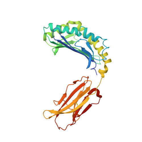
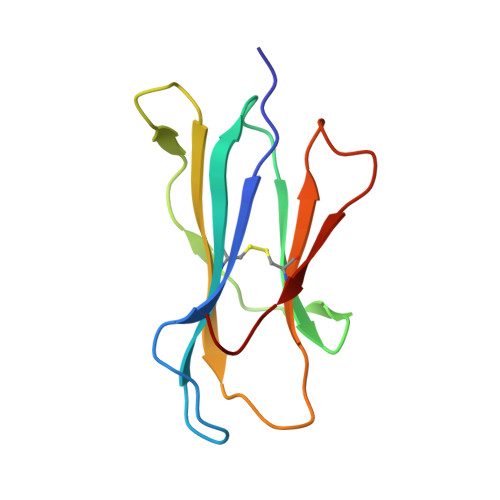
The crystal structure of avian CD1 reveals a smaller, more primordial antigen-binding pocket compared to mammalian CD1

Zajonc, D.M.Striegl, H.Dascher, C.C.Wilson, I.A.

(2008) Proc Natl Acad Sci U S A 105: 17925-17930

  • DOI: https://doi.org/10.1073/pnas.0809814105
  • Primary Citation of Related Structures:  
    3DBX

  • PubMed Abstract: 

    The molecular details of glycolipid presentation by CD1 antigen-presenting molecules are well studied in mammalian systems. However, little is known about how these non-classical MHC class I (MHCI) molecules diverged from the MHC locus to create a more complex, hydrophobic binding groove that binds lipids rather than peptides. To address this fundamental question, we have determined the crystal structure of an avian CD1 (chCD1-2) that shares common ancestry with mammalian CD1 from approximately 310 million years ago. The chCD1-2 antigen-binding site consists of a compact, narrow, central hydrophobic groove or pore rather than the more open, 2-pocket architecture observed in mammalian CD1s. Potential antigens then would be restricted in size to single-chain lipids or glycolipids. An endogenous ligand, possibly palmitic acid, serves to illuminate the mode and mechanism of ligand interaction with chCD1-2. The palmitate alkyl chain is inserted into the relatively shallow hydrophobic pore; its carboxyl group emerges at the receptor surface and is stabilized by electrostatic and hydrogen bond interactions with an arginine residue that is conserved in all known CD1 proteins. In addition, other novel features, such as an A' loop that interrupts and segments the normally long, continuous alpha1 helix, are unique to chCD1-2 and contribute to the unusually narrow binding groove, thereby limiting its size. Because birds and mammals share a common ancestor, but the rate of evolution is slower in birds than in mammals, the chCD1-2-binding groove probably represents a more primordial CD1-binding groove.


  • Organizational Affiliation

    Division of Cell Biology, La Jolla Institute for Allergy and Immunology, 9420 Athena Circle, La Jolla, CA 92037, USA. dzajonc@liai.org


Macromolecules
Find similar proteins by:  (by identity cutoff)  |  3D Structure
Entity ID: 1
MoleculeChains Sequence LengthOrganismDetailsImage
CD1-2 antigen289Gallus gallusMutation(s): 0 
Gene Names: CD1-2
UniProt
Find proteins for Q5GL29 (Gallus gallus)
Explore Q5GL29 
Go to UniProtKB:  Q5GL29
Entity Groups  
Sequence Clusters30% Identity50% Identity70% Identity90% Identity95% Identity100% Identity
UniProt GroupQ5GL29
Sequence Annotations
Expand
  • Reference Sequence
Find similar proteins by:  (by identity cutoff)  |  3D Structure
Entity ID: 2
MoleculeChains Sequence LengthOrganismDetailsImage
Beta-2-microglobulin99Homo sapiensMutation(s): 0 
Gene Names: B2MCDABP0092HDCMA22P
UniProt & NIH Common Fund Data Resources
Find proteins for P61769 (Homo sapiens)
Explore P61769 
Go to UniProtKB:  P61769
PHAROS:  P61769
GTEx:  ENSG00000166710 
Entity Groups  
Sequence Clusters30% Identity50% Identity70% Identity90% Identity95% Identity100% Identity
UniProt GroupP61769
Sequence Annotations
Expand
  • Reference Sequence
Experimental Data & Validation

Experimental Data

  • Method: X-RAY DIFFRACTION
  • Resolution: 2.00 Å
  • R-Value Free: 0.266 
  • R-Value Work: 0.216 
  • R-Value Observed: 0.218 
  • Space Group: P 41 21 2
Unit Cell:
Length ( Å )Angle ( ˚ )
a = 92.154α = 90
b = 92.154β = 90
c = 96.693γ = 90
Software Package:
Software NamePurpose
REFMACrefinement
HKL-2000data collection
DENZOdata reduction
SCALEPACKdata scaling
MOLREPphasing

Structure Validation

View Full Validation Report


Ligand Structure Quality Assessment 


Entry History 

Deposition Data

Revision History  (Full details and data files)

  • Version 1.0: 2008-11-25
    Type: Initial release
  • Version 1.1: 2011-07-13
    Changes: Non-polymer description, Version format compliance
  • Version 1.2: 2020-07-29
    Type: Remediation
    Reason: Carbohydrate remediation
    Changes: Data collection, Database references, Derived calculations, Structure summary
  • Version 1.3: 2023-11-01
    Changes: Data collection, Database references, Refinement description, Structure summary