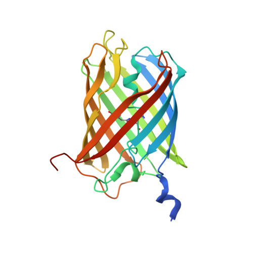
Crystal structure and Raman studies of dsFP483, a cyan fluorescent protein from Discosoma striata.
Malo, G.D., Wang, M., Wu, D., Stelling, A.L., Tonge, P.J., Wachter, R.M.(2008) J Mol Biology 378: 869-884
- PubMed: 18395223 Search on PubMedSearch on PubMed Central
- DOI: https://doi.org/10.1016/j.jmb.2008.02.069
- Primary Citation Related Structures:
3CGL - PubMed Abstract:
To better understand the diverse mechanisms of spectral tuning operational in fluorescent proteins (FPs), we determined the 2.1-A X-ray structure of dsFP483 from the reef-building coral Discosoma. This protein is a member of the cyan class of Anthozoa FPs and exhibits broad, double-humped excitation and absorbance bands, with a maximum at 437-440 nm and a shoulder at 453 nm. Although these features support a heterogeneous ground state for the protein-intrinsic chromophore, peak fluorescence occurs at 483 nm for all excitation wavelengths, suggesting a common emissive state. Optical properties are insensitive to changes in pH over the entire range of protein stability. The refined crystal structure of the biological tetramer (space group C2) demonstrates that all protomers bear a cis-coplanar chromophore chemically identical with that in green fluorescent protein (GFP). To test the roles of specific residues in color modulation, we investigated the optical properties of the H163Q and K70M variants. Although absorbance bands remain broad, peak excitation maxima are red shifted to 455 and 460 nm, emitting cyan light and green light, respectively. To probe chromophore ground-state features, we collected Raman spectra using 752-nm excitation. Surprisingly, the positions of key Raman bands of wild-type dsFP483 are most similar to those of the neutral GFP chromophore, whereas the K70M spectra are more closely aligned with the anionic form. The Raman data provide further evidence of a mixed ground state with chromophore populations that are modulated by mutation. Possible internal protonation equilibria, structural heterogeneity in the binding sites, and excited-state proton transfer mechanisms are discussed. Structural alignments of dsFP483 with the homologs DsRed, amFP486, and zFP538-K66M suggest that natural selection for cyan is an exquisitely fine-tuned and highly cooperative process involving a network of electrostatic interactions that may vary substantially in composition and arrangement.
- Department of Chemistry and Biochemistry, Arizona State University, Tempe, AZ 85287-1604, USA.
Organizational Affiliation: