Structural insights into the modulation of the redox properties of two Geobacter sulfurreducens homologous triheme cytochromes.
Morgado, L., Bruix, M., Orshonsky, V., Londer, Y.Y., Duke, N.E., Yang, X., Pokkuluri, P.R., Schiffer, M., Salgueiro, C.A.(2008) Biochim Biophys Acta 1777: 1157-1165
- PubMed: 18534185 Search on PubMed
- DOI: https://doi.org/10.1016/j.bbabio.2008.04.043
- Primary Citation Related Structures:
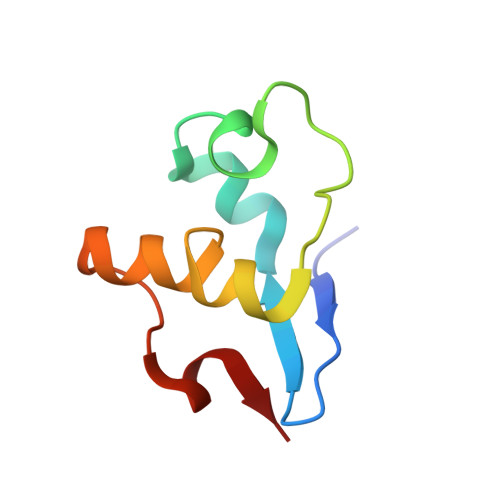
3BXU - PubMed Abstract:
The redox properties of a periplasmic triheme cytochrome, PpcB from Geobacter sulfurreducens, were studied by NMR and visible spectroscopy. The structure of PpcB was determined by X-ray diffraction. PpcB is homologous to PpcA (77% sequence identity), which mediates cytoplasmic electron transfer to extracellular acceptors and is crucial in the bioenergetic metabolism of Geobacter spp. The heme core structure of PpcB in solution, probed by 2D-NMR, was compared to that of PpcA. The results showed that the heme core structures of PpcB and PpcA in solution are similar, in contrast to their crystal structures where the heme cores of the two proteins differ from each other. NMR redox titrations were carried out for both proteins and the order of oxidation of the heme groups was determined. The microscopic properties of PpcB and PpcA redox centers showed important differences: (i) the order in which hemes become oxidized is III-I-IV for PpcB, as opposed to I-IV-III for PpcA; (ii) the redox-Bohr effect is also different in the two proteins. The different redox features observed between PpcB and PpcA suggest that each protein uniquely modulates the properties of their co-factors to assure effectiveness in their respective metabolic pathways. The origins of the observed differences are discussed.
- Requimte-CQFB, Departamento de Química, Faculdade de Ciências e Tecnologia, Universidade Nova de Lisboa, Campus Caparica, 2829-516 Caparica, Portugal.
Organizational Affiliation: