3BUO

Crystal structure of c-Cbl-TKB domain complexed with its binding motif in EGF receptor'


Experimental Data Snapshot

  • Method: X-RAY DIFFRACTION
  • Resolution: 2.60 Å
  • R-Value Free: 0.278 
  • R-Value Work: 0.231 

wwPDB Validation   3D Report Full Report


This is version 1.6 of the entry. See complete history


Literature

Structural basis for a novel intrapeptidyl H-bond and reverse binding of c-Cbl-TKB domain substrates

Ng, C.Jackson, R.A.Buschdorf, J.P.Sun, Q.Guy, G.R.Sivaraman, J.

(2008) EMBO J 27: 804-816

  • DOI: https://doi.org/10.1038/emboj.2008.18
  • Primary Citation of Related Structures:  
    3BUM, 3BUN, 3BUO, 3BUW, 3BUX

  • PubMed Abstract: 

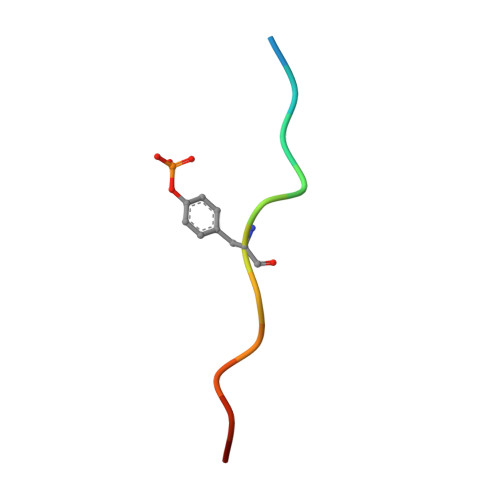
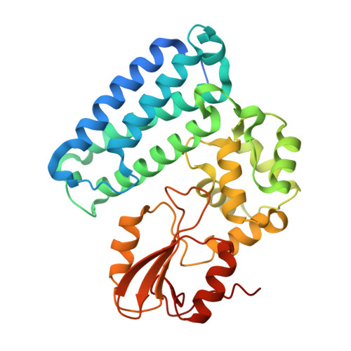
    The c-Cbl tyrosine kinase binding domain (Cbl-TKB), essentially an 'embedded' SH2 domain, has a critical role in targeting proteins for ubiquitination. To address how this domain can bind to disparate recognition mofits and to determine whether this results in variations in substrate-binding affinity, we compared crystal structures of the Cbl-TKB domain complexed with phosphorylated peptides of Sprouty2, Sprouty4, epidermal growth factor receptor, Syk, and c-Met receptors and validated the binding with point-mutational analyses using full-length proteins. An obligatory, intrapeptidyl H-bond between the phosphotyrosine and the conserved asparagine or adjacent arginine is essential for binding and orients the peptide into a positively charged pocket on c-Cbl. Surprisingly, c-Met bound to Cbl in the reverse direction, which is unprecedented for SH2 domain binding. The necessity of this intrapeptidyl H-bond was confirmed with isothermal titration calorimetry experiments that also showed Sprouty2 to have the highest binding affinity to c-Cbl; this may enable the selective sequestration of c-Cbl from other target proteins.


  • Organizational Affiliation

    Department of Biological Sciences, National University of Singapore, Singapore.


Macromolecules

Find similar proteins by:  Sequence   |   3D Structure  

Entity ID: 1
MoleculeChains Sequence LengthOrganismDetailsImage
13-meric peptide from Epidermal growth factor receptor
A, C
13Homo sapiensMutation(s): 0 
UniProt & NIH Common Fund Data Resources
Find proteins for P00533 (Homo sapiens)
Explore P00533 
Go to UniProtKB:  P00533
PHAROS:  P00533
GTEx:  ENSG00000146648 
Entity Groups  
Sequence Clusters30% Identity50% Identity70% Identity90% Identity95% Identity100% Identity
UniProt GroupP00533
Sequence Annotations
Expand
  • Reference Sequence
Find similar proteins by:  (by identity cutoff)  |  3D Structure
Entity ID: 2
MoleculeChains Sequence LengthOrganismDetailsImage
E3 ubiquitin-protein ligase CBL
B, D
329Homo sapiensMutation(s): 0 
Gene Names: CBLCBL2RNF55
EC: 6.3.2
UniProt & NIH Common Fund Data Resources
Find proteins for P22681 (Homo sapiens)
Explore P22681 
Go to UniProtKB:  P22681
PHAROS:  P22681
GTEx:  ENSG00000110395 
Entity Groups  
Sequence Clusters30% Identity50% Identity70% Identity90% Identity95% Identity100% Identity
UniProt GroupP22681
Sequence Annotations
Expand
  • Reference Sequence
Small Molecules
Modified Residues  1 Unique
IDChains TypeFormula2D DiagramParent
PTR
Query on PTR
A, C
L-PEPTIDE LINKINGC9 H12 N O6 PTYR
Experimental Data & Validation

Experimental Data

  • Method: X-RAY DIFFRACTION
  • Resolution: 2.60 Å
  • R-Value Free: 0.278 
  • R-Value Work: 0.231 
  • Space Group: P 1 21 1
Unit Cell:
Length ( Å )Angle ( ˚ )
a = 63.862α = 90
b = 110.173β = 89.94
c = 55.821γ = 90
Software Package:
Software NamePurpose
DENZOdata reduction
SCALEPACKdata scaling
MOLREPphasing
CNSrefinement
PDB_EXTRACTdata extraction
HKL-2000data reduction
HKL-2000data scaling

Structure Validation

View Full Validation Report


Entry History 

Deposition Data

Revision History  (Full details and data files)

  • Version 1.0: 2008-02-26
    Type: Initial release
  • Version 1.1: 2011-07-13
    Changes: Version format compliance
  • Version 1.2: 2015-01-14
    Changes: Derived calculations
  • Version 1.3: 2017-10-25
    Changes: Refinement description
  • Version 1.4: 2018-09-19
    Changes: Data collection, Source and taxonomy
  • Version 1.5: 2023-11-01
    Changes: Data collection, Database references, Derived calculations, Refinement description
  • Version 1.6: 2023-11-15
    Changes: Data collection