3AQY | pdb_00003aqy

Crystal structure of Plodia interpunctella beta-GRP/GNBP3 N-terminal domain


Experimental Data Snapshot

  • Method: X-RAY DIFFRACTION
  • Resolution: 1.58 Å
  • R-Value Free: 
    0.238 (Depositor), 0.237 (DCC) 
  • R-Value Work: 
    0.207 (Depositor), 0.207 (DCC) 
  • R-Value Observed: 
    0.209 (Depositor) 

Starting Model: experimental
View more details

wwPDB Validation 3D Report Full Report

Validation slider image for 3AQY

This is version 1.3 of the entry. See complete history

Literature

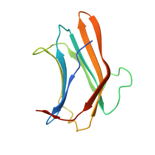
Structural insights into recognition of triple-helical beta-glucans by an insect fungal receptor

Kanagawa, M.Satoh, T.Ikeda, A.Adachi, Y.Ohno, N.Yamaguchi, Y.

(2011) J Biological Chem 286: 29158-29165

  • DOI: https://doi.org/10.1074/jbc.M111.256701
  • Primary Citation Related Structures: 
    3AQX, 3AQY, 3AQZ

  • PubMed Abstract: 

    The innate ability to detect pathogens is achieved by pattern recognition receptors, which recognize non-self-components such as β1,3-glucan. β1,3-Glucans form a triple-helical structure stabilized by interchain hydrogen bonds. β1,3-Glucan recognition protein (βGRP)/gram-negative bacteria-binding protein 3 (GNBP3), one of the pattern recognition receptors, binds to long, structured β1,3-glucan to initiate innate immune response. However, binding details and how specificity is achieved in such receptors remain important unresolved issues. We solved the crystal structures of the N-terminal β1,3-glucan recognition domain of βGRP/GNBP3 (βGRP-N) in complex with the β1,3-linked glucose hexamer, laminarihexaose. In the crystals, three structured laminarihexaoses simultaneously interact through six glucose residues (two from each chain) with one βGRP-N. The spatial arrangement of the laminarihexaoses bound to βGRP-N is almost identical to that of a β1,3-glucan triple-helical structure. Therefore, our crystallographic structures together with site-directed mutagenesis data provide a structural basis for the unique recognition by such receptors of the triple-helical structure of β1,3-glucan.


  • Organizational Affiliation
    • Structural Glycobiology Team, Systems Glycobiology Research Group, Chemical Biology Department, RIKEN, Advanced Science Institute, 2-1 Hirosawa Wako, Saitama 351-0198, Japan and.

Macromolecule Content 

  • Total Structure Weight: 24.35 kDa 
  • Atom Count: 1,801 
  • Modeled Residue Count: 198 
  • Deposited Residue Count: 212 
  • Unique protein chains: 1

Macromolecules

Find similar proteins by:|  3D Structure
Entity ID: 1
MoleculeChains  Sequence LengthOrganismDetailsImage
Beta-1,3-glucan-binding protein
A, B
106Plodia interpunctellaMutation(s): 0 
UniProt
Find proteins for Q8MU95 (Plodia interpunctella)
Explore Q8MU95 
Go to UniProtKB:  Q8MU95
Entity Groups
Sequence Clusters30% Identity50% Identity70% Identity90% Identity95% Identity100% Identity
UniProt GroupQ8MU95
Sequence Annotations
Expand
Reference Sequence

Experimental Data & Validation

Experimental Data

  • Method: X-RAY DIFFRACTION
  • Resolution: 1.58 Å
  • R-Value Free:  0.238 (Depositor), 0.237 (DCC) 
  • R-Value Work:  0.207 (Depositor), 0.207 (DCC) 
  • R-Value Observed: 0.209 (Depositor) 
Space Group: C 1 2 1
Unit Cell:
Length ( Å )Angle ( ˚ )
a = 106.35α = 90
b = 38.034β = 117.34
c = 55.63γ = 90
Software Package:
Software NamePurpose
HKL-2000data collection
MOLREPphasing
REFMACrefinement
DENZOdata reduction
SCALEPACKdata scaling

Structure Validation

View Full Validation Report


Entry History 

Deposition Data

Revision History  (Full details and data files)

  • Version 1.0: 2011-06-22
    Type: Initial release
  • Version 1.1: 2011-07-13
    Changes: Version format compliance
  • Version 1.2: 2013-06-19
    Changes: Database references
  • Version 1.3: 2023-11-01
    Changes: Data collection, Database references, Refinement description