3ALA | pdb_00003ala

Crystal structure of vascular adhesion protein-1 in space group C2


Experimental Data Snapshot

  • Method: X-RAY DIFFRACTION
  • Resolution: 2.90 Å
  • R-Value Free: 
    0.286 (Depositor), 0.282 (DCC) 
  • R-Value Work: 
    0.250 (Depositor), 0.249 (DCC) 
  • R-Value Observed: 
    0.252 (Depositor) 

Starting Model: experimental
View more details

wwPDB Validation 3D Report Full Report

Validation slider image for 3ALA

Ligand Structure Quality Assessment 


This is version 2.1 of the entry. See complete history

Literature

A new crystal form of human vascular adhesion protein 1

Ernberg, K.McGrath, A.P.Peat, T.S.Adams, T.E.Xiao, X.Pham, T.Newman, J.McDonald, I.A.Collyer, C.A.Guss, J.M.

(2010) Acta Crystallogr Sect F Struct Biol Cryst Commun 66: 1572-1578

  • DOI: https://doi.org/10.1107/S1744309110041515
  • Primary Citation Related Structures: 
    3ALA

  • PubMed Abstract: 

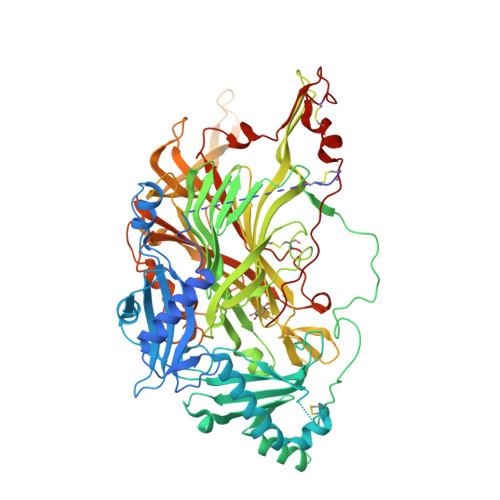
    Human vascular adhesion protein 1 (VAP-1) is involved in lymphocyte-endothelial cell adhesion and has been implicated in many human inflammatory diseases. VAP-1 is a member of the copper amine oxidase family of enzymes with a trihydroxyphenylalanine quinone (TPQ) cofactor. Previously characterized crystals of VAP-1 suffered from anisotropy and contained disordered regions; in addition, one form was consistently twinned. In an effort to grow crystals that diffracted to higher resolution for inhibitor-binding studies, a construct with an N-terminal deletion was made and expressed in the Chinese hamster ovary (CHO) glycosylation mutant cell line Lec8. Screening produced crystals that displayed some anisotropy and contained seven molecules per asymmetric unit. These crystals belonged to space group C2, with unit-cell parameters a=394.5, b=115.8, c=179.3 Å, β=112.3°. The structure was refined to a resolution of 2.9 Å, with Rcryst and Rfree values of 0.250 and 0.286, respectively.


  • Organizational Affiliation
    • School of Molecular Bioscience, University of Sydney, NSW 2006, Australia.

Macromolecule Content 

  • Total Structure Weight: 595.58 kDa 
  • Atom Count: 39,103 
  • Modeled Residue Count: 4,939 
  • Deposited Residue Count: 5,236 
  • Unique protein chains: 1

Macromolecules

Find similar proteins by:|  3D Structure
Entity ID: 1
MoleculeChains  Sequence LengthOrganismDetailsImage
Membrane primary amine oxidase
A, B, C, D, E
A, B, C, D, E, F, G
748Homo sapiensMutation(s): 0 
Gene Names: AOC3VAP1
EC: 1.4.3.21
UniProt & NIH Common Fund Data Resources
Find proteins for Q16853 (Homo sapiens)
Explore Q16853 
Go to UniProtKB:  Q16853
PHAROS:  Q16853
GTEx:  ENSG00000131471 
Entity Groups
Sequence Clusters30% Identity50% Identity70% Identity90% Identity95% Identity100% Identity
UniProt GroupQ16853
Glycosylation
Glycosylation Sites: 3Go to GlyGen: Q16853-1
Sequence Annotations
Expand
Reference Sequence

Oligosaccharides

Help  
Entity ID: 2
MoleculeChains Length2D Diagram GlycosylationD Interactions
beta-D-mannopyranose-(1-4)-2-acetamido-2-deoxy-beta-D-glucopyranose-(1-4)-2-acetamido-2-deoxy-beta-D-glucopyranose
H, K, M, P, S
H, K, M, P, S, V, Y
3N-Glycosylation
Glycosylation Resources
GlyTouCan: G15407YE
GlyCosmos: G15407YE
GlyGen: G15407YE
Entity ID: 3
MoleculeChains Length2D Diagram GlycosylationD Interactions
2-acetamido-2-deoxy-beta-D-glucopyranose-(1-4)-2-acetamido-2-deoxy-beta-D-glucopyranose
I, N, Q, T, W
I, N, Q, T, W, Z
2N-Glycosylation
Glycosylation Resources
GlyTouCan: G42666HT
GlyCosmos: G42666HT
GlyGen: G42666HT
Entity ID: 4
MoleculeChains Length2D Diagram GlycosylationD Interactions
beta-L-fucopyranose-(1-6)-2-acetamido-2-deoxy-beta-D-glucopyranose
J, O, R, U, X
2N-Glycosylation
Glycosylation Resources
GlyTouCan: G61843VN
GlyCosmos: G61843VN
GlyGen: G61843VN
Entity ID: 5
MoleculeChains Length2D Diagram GlycosylationD Interactions
2-acetamido-2-deoxy-beta-D-glucopyranose-(1-4)-[beta-L-fucopyranose-(1-6)]2-acetamido-2-deoxy-beta-D-glucopyranose
L
3N-Glycosylation
Glycosylation Resources
GlyTouCan: G28454KX
GlyCosmos: G28454KX
GlyGen: G28454KX
Entity ID: 6
MoleculeChains Length2D Diagram GlycosylationD Interactions
alpha-L-fucopyranose-(1-6)-2-acetamido-2-deoxy-beta-D-glucopyranoseAA [auth a]2N-Glycosylation
Glycosylation Resources
GlyTouCan: G86851RC
GlyCosmos: G86851RC
GlyGen: G86851RC

Small Molecules

Ligands 4 Unique
IDChains Name / Formula / InChI Key2D Diagram3D Interactions
NAG

Query on NAG


Download:Ideal Coordinates CCD File
IA [auth B],
PA [auth D]
2-acetamido-2-deoxy-beta-D-glucopyranose
C8 H15 N O6
OVRNDRQMDRJTHS-FMDGEEDCSA-N
GOL

Query on GOL


Download:Ideal Coordinates CCD File
EA [auth A]GLYCEROL
C3 H8 O3
PEDCQBHIVMGVHV-UHFFFAOYSA-N
CU

Query on CU


Download:Ideal Coordinates CCD File
BA [auth A]
FA [auth B]
JA [auth C]
MA [auth D]
QA [auth E]
BA [auth A],
FA [auth B],
JA [auth C],
MA [auth D],
QA [auth E],
TA [auth F],
WA [auth G]
COPPER (II) ION
Cu
JPVYNHNXODAKFH-UHFFFAOYSA-N
CA

Query on CA


Download:Ideal Coordinates CCD File
CA [auth A]
DA [auth A]
GA [auth B]
HA [auth B]
KA [auth C]
CA [auth A],
DA [auth A],
GA [auth B],
HA [auth B],
KA [auth C],
LA [auth C],
NA [auth D],
OA [auth D],
RA [auth E],
SA [auth E],
UA [auth F],
VA [auth F],
XA [auth G],
YA [auth G]
CALCIUM ION
Ca
BHPQYMZQTOCNFJ-UHFFFAOYSA-N
Modified Residues  1 Unique
IDChains TypeFormula2D DiagramParent
TPQ
Query on TPQ
A, B, C, D, E
A, B, C, D, E, F, G
L-PEPTIDE LINKINGC9 H9 N O5TYR

Experimental Data & Validation

Experimental Data

  • Method: X-RAY DIFFRACTION
  • Resolution: 2.90 Å
  • R-Value Free:  0.286 (Depositor), 0.282 (DCC) 
  • R-Value Work:  0.250 (Depositor), 0.249 (DCC) 
  • R-Value Observed: 0.252 (Depositor) 
Space Group: C 1 2 1
Unit Cell:
Length ( Å )Angle ( ˚ )
a = 394.468α = 90
b = 115.826β = 112.34
c = 179.286γ = 90
Software Package:
Software NamePurpose
SCALAdata scaling
REFMACrefinement
PDB_EXTRACTdata extraction
MOSFLMdata reduction
PHASERphasing

Structure Validation

View Full Validation Report


Ligand Structure Quality Assessment 


Entry History 

Deposition Data

Revision History  (Full details and data files)

  • Version 1.0: 2010-12-15
    Type: Initial release
  • Version 1.1: 2011-07-13
    Changes: Version format compliance
  • Version 1.2: 2012-04-25
    Changes: Database references
  • Version 2.0: 2020-07-29
    Type: Remediation
    Reason: Carbohydrate remediation
    Changes: Advisory, Atomic model, Data collection, Database references, Derived calculations, Non-polymer description, Structure summary
  • Version 2.1: 2023-11-01
    Changes: Advisory, Data collection, Database references, Refinement description, Structure summary