2YCF

Crystal Structure of Checkpoint Kinase 2 in complex with Inhibitor PV1531


Experimental Data Snapshot

  • Method: X-RAY DIFFRACTION
  • Resolution: 1.77 Å
  • R-Value Free: 0.218 
  • R-Value Work: 0.196 
  • R-Value Observed: 0.197 

wwPDB Validation   3D Report Full Report


Ligand Structure Quality Assessment 


This is version 1.1 of the entry. See complete history


Literature

Structural Characterization of Inhibitor Complexes with Checkpoint Kinase 2 (Chk2), a Drug Target for Cancer Therapy.

Lountos, G.T.Jobson, A.G.Tropea, J.E.Self, C.R.Zhang, G.Pommier, Y.Shoemaker, R.H.Waugh, D.S.

(2011) J Struct Biol 176: 292

  • DOI: https://doi.org/10.1016/j.jsb.2011.09.008
  • Primary Citation of Related Structures:  
    2XK9, 2YCF, 2YCQ, 2YCR, 2YCS

  • PubMed Abstract: 

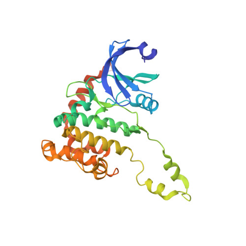
    Chk2 (checkpoint kinase 2) is a serine/threonine kinase that participates in a series of signaling networks responsible for maintaining genomic integrity and responding to DNA damage. The development of selective Chk2 inhibitors has recently attracted much interest as a means of sensitizing cancer cells to current DNA-damaging agents used in the treatment of cancer. Additionally, selective Chk2 inhibitors may reduce p53-mediated apoptosis in normal tissues, thereby helping to mitigate adverse side effects from chemotherapy and radiation. Thus far, relatively few selective inhibitors of Chk2 have been described and none have yet progressed into clinical trials. Here, we report crystal structures of the catalytic domain of Chk2 in complex with a novel series of potent and selective small molecule inhibitors. These compounds exhibit nanomolar potencies and are selective for Chk2 over Chk1. The structures reported here elucidate the binding modes of these inhibitors to Chk2 and provide information that can be exploited for the structure-assisted design of novel chemotherapeutics.


  • Organizational Affiliation

    Center for Cancer Research, National Cancer Institute at Frederick, Frederick, MD 21702-1201, USA.


Macromolecules
Find similar proteins by:  (by identity cutoff)  |  3D Structure
Entity ID: 1
MoleculeChains Sequence LengthOrganismDetailsImage
SERINE/THREONINE-PROTEIN KINASE CHK2322Homo sapiensMutation(s): 0 
EC: 2.7.11.1
UniProt & NIH Common Fund Data Resources
Find proteins for O96017 (Homo sapiens)
Explore O96017 
Go to UniProtKB:  O96017
PHAROS:  O96017
GTEx:  ENSG00000183765 
Entity Groups  
Sequence Clusters30% Identity50% Identity70% Identity90% Identity95% Identity100% Identity
UniProt GroupO96017
Sequence Annotations
Expand
  • Reference Sequence
Small Molecules
Ligands 3 Unique
IDChains Name / Formula / InChI Key2D Diagram3D Interactions
YCF
Query on YCF

Download Ideal Coordinates CCD File 
B [auth A](2E)-N-hydroxy-2-[1-(4-{[(4-{(1E)-1-[2-(N'-hydroxycarbamimidoyl)hydrazinylidene]ethyl}phenyl)carbamoyl]amino}phenyl)ethylidene]hydrazinecarboximidamide
C19 H24 N10 O3
VVGIJVMXIRYKIO-DHSUVYDUSA-N
NO3
Query on NO3

Download Ideal Coordinates CCD File 
D [auth A]NITRATE ION
N O3
NHNBFGGVMKEFGY-UHFFFAOYSA-N
MG
Query on MG

Download Ideal Coordinates CCD File 
C [auth A]MAGNESIUM ION
Mg
JLVVSXFLKOJNIY-UHFFFAOYSA-N
Binding Affinity Annotations 
IDSourceBinding Affinity
YCF PDBBind:  2YCF IC50: 0.03 (nM) from 1 assay(s)
Binding MOAD:  2YCF IC50: 0.03 (nM) from 1 assay(s)
Experimental Data & Validation

Experimental Data

  • Method: X-RAY DIFFRACTION
  • Resolution: 1.77 Å
  • R-Value Free: 0.218 
  • R-Value Work: 0.196 
  • R-Value Observed: 0.197 
  • Space Group: P 32 2 1
Unit Cell:
Length ( Å )Angle ( ˚ )
a = 90.465α = 90
b = 90.465β = 90
c = 93.639γ = 120
Software Package:
Software NamePurpose
REFMACrefinement
HKL-3000data reduction
SCALEPACKdata scaling
MOLREPphasing

Structure Validation

View Full Validation Report


Ligand Structure Quality Assessment 


Entry History 

Deposition Data

Revision History  (Full details and data files)

  • Version 1.0: 2011-11-16
    Type: Initial release
  • Version 1.1: 2023-12-20
    Changes: Data collection, Database references, Derived calculations, Other, Refinement description