2Y5W

Crystal structure of Drosophila melanogaster kinesin-1 motor domain dimer


Experimental Data Snapshot

  • Method: X-RAY DIFFRACTION
  • Resolution: 2.70 Å
  • R-Value Free: 0.289 
  • R-Value Work: 0.210 
  • R-Value Observed: 0.214 

wwPDB Validation   3D Report Full Report


Ligand Structure Quality Assessment 


This is version 1.2 of the entry. See complete history


Literature

The Structure of the Kinesin-1 Motor-Tail Complex Reveals the Mechanism of Autoinhibition.

Kaan, H.Y.K.Hackney, D.D.Kozielski, F.

(2011) Science 333: 883

  • DOI: https://doi.org/10.1126/science.1204824
  • Primary Citation of Related Structures:  
    2Y5W, 2Y65

  • PubMed Abstract: 

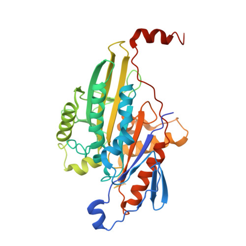
    When not transporting cargo, kinesin-1 is autoinhibited by binding of a tail region to the motor domains, but the mechanism of inhibition is unclear. We report the crystal structure of a motor domain dimer in complex with its tail domain at 2.2 angstroms and compare it with a structure of the motor domain alone at 2.7 angstroms. These structures indicate that neither an induced conformational change nor steric blocking is the cause of inhibition. Instead, the tail cross-links the motor domains at a second position, in addition to the coiled coil. This "double lockdown," by cross-linking at two positions, prevents the movement of the motor domains that is needed to undock the neck linker and release adenosine diphosphate. This autoinhibition mechanism could extend to some other kinesins.


  • Organizational Affiliation

    The Beatson Institute for Cancer Research, Switchback Road, Bearsden, Glasgow G61 1BD, Scotland, UK.


Macromolecules
Find similar proteins by:  (by identity cutoff)  |  3D Structure
Entity ID: 1
MoleculeChains Sequence LengthOrganismDetailsImage
KINESIN HEAVY CHAIN
A, B
365Drosophila melanogasterMutation(s): 0 
UniProt
Find proteins for P17210 (Drosophila melanogaster)
Explore P17210 
Go to UniProtKB:  P17210
Entity Groups  
Sequence Clusters30% Identity50% Identity70% Identity90% Identity95% Identity100% Identity
UniProt GroupP17210
Sequence Annotations
Expand
  • Reference Sequence
Experimental Data & Validation

Experimental Data

  • Method: X-RAY DIFFRACTION
  • Resolution: 2.70 Å
  • R-Value Free: 0.289 
  • R-Value Work: 0.210 
  • R-Value Observed: 0.214 
  • Space Group: P 21 21 21
Unit Cell:
Length ( Å )Angle ( ˚ )
a = 76.89α = 90
b = 89.18β = 90
c = 138.35γ = 90
Software Package:
Software NamePurpose
REFMACrefinement
iMOSFLMdata reduction
SCALAdata scaling
PHASERphasing

Structure Validation

View Full Validation Report


Ligand Structure Quality Assessment 


Entry History 

Deposition Data

Revision History  (Full details and data files)

  • Version 1.0: 2011-08-24
    Type: Initial release
  • Version 1.1: 2011-08-31
    Changes: Structure summary
  • Version 1.2: 2023-12-20
    Changes: Data collection, Database references, Derived calculations, Other, Refinement description