2VNL

MUTANT Y108Wdel OF THE HEADBINDING DOMAIN OF PHAGE P22 TAILSPIKE C- TERMINally fused to ISOLEUCINE ZIPPER pIIGCN4 (chimera II)


Experimental Data Snapshot

  • Method: X-RAY DIFFRACTION
  • Resolution: 1.80 Å
  • R-Value Free: 0.229 
  • R-Value Work: 0.190 
  • R-Value Observed: 0.192 

wwPDB Validation   3D Report Full Report


This is version 1.5 of the entry. See complete history


Literature

Bacteriophage P22 Tailspike: Structure of the Complete Protein and Function of the Interdomain Linker

Seul, A.Mueller, J.J.Andres, D.Stettner, E.Heinemann, U.Seckler, R.

(2014) Acta Crystallogr D Biol Crystallogr 70: 1336

  • DOI: https://doi.org/10.1107/S1399004714002685
  • Primary Citation of Related Structures:  
    2VKY, 2VNL, 2XC1

  • PubMed Abstract: 

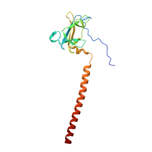
    Attachment of phages to host cells, followed by phage DNA ejection, represents the first stage of viral infection of bacteria. Salmonella phage P22 has been extensively studied, serving as an experimental model for bacterial infection by phages. P22 engages bacteria by binding to the sugar moiety of lipopolysaccharides using the viral tailspike protein for attachment. While the structures of the N-terminal particle-binding domain and the major receptor-binding domain of the tailspike have been analyzed individually, the three-dimensional organization of the intact protein, including the highly conserved linker region between the two domains, remained unknown. A single amino-acid exchange in the linker sequence made it possible to crystallize the full-length protein. Two crystal structures of the linker region are presented: one attached to the N-terminal domain and the other present within the complete tailspike protein. Both retain their biological function, but the mutated full-length tailspike displays a retarded folding pathway. Fitting of the full-length tailspike into a published cryo-electron microscopy map of the P22 virion requires an elastic distortion of the crystal structure. The conservation of the linker suggests a role in signal transmission from the distal tip of the molecule to the phage head, eventually leading to DNA ejection.


  • Organizational Affiliation

    Physikalische Biochemie, Universität Potsdam, Karl-Liebknecht-Strasse 24-25, 14476 Potsdam, Germany.


Macromolecules
Find similar proteins by:  (by identity cutoff)  |  3D Structure
Entity ID: 1
MoleculeChains Sequence LengthOrganismDetailsImage
BIFUNCTIONAL TAIL PROTEIN, PIIGCN4151Lederbergvirus P22Saccharomyces cerevisiae
This entity is chimeric
Mutation(s): 1 
UniProt
Find proteins for P03069 (Saccharomyces cerevisiae (strain ATCC 204508 / S288c))
Explore P03069 
Go to UniProtKB:  P03069
Find proteins for P12528 (Salmonella phage P22)
Explore P12528 
Go to UniProtKB:  P12528
Entity Groups  
Sequence Clusters30% Identity50% Identity70% Identity90% Identity95% Identity100% Identity
UniProt GroupsP03069P12528
Sequence Annotations
Expand
  • Reference Sequence
Experimental Data & Validation

Experimental Data

  • Method: X-RAY DIFFRACTION
  • Resolution: 1.80 Å
  • R-Value Free: 0.229 
  • R-Value Work: 0.190 
  • R-Value Observed: 0.192 
  • Space Group: H 3
Unit Cell:
Length ( Å )Angle ( ˚ )
a = 58.012α = 90
b = 58.012β = 90
c = 156.11γ = 120
Software Package:
Software NamePurpose
REFMACrefinement
XDSdata reduction
XSCALEdata scaling
PHASERphasing

Structure Validation

View Full Validation Report


Entry History 

Deposition Data

Revision History  (Full details and data files)

  • Version 1.0: 2009-02-10
    Type: Initial release
  • Version 1.1: 2014-05-14
    Changes: Database references, Derived calculations, Non-polymer description, Other, Structure summary, Version format compliance
  • Version 1.2: 2014-05-21
    Changes: Database references
  • Version 1.3: 2017-03-15
    Changes: Source and taxonomy
  • Version 1.4: 2019-01-30
    Changes: Data collection, Experimental preparation
  • Version 1.5: 2023-12-13
    Changes: Data collection, Database references, Derived calculations, Other, Refinement description