2VDN | pdb_00002vdn

Re-refinement of Integrin AlphaIIbBeta3 Headpiece Bound to Antagonist Eptifibatide


Experimental Data Snapshot

  • Method: X-RAY DIFFRACTION
  • Resolution: 2.90 Å
  • R-Value Free: 
    0.213 (Depositor), 0.216 (DCC) 
  • R-Value Work: 
    0.163 (Depositor), 0.170 (DCC) 
  • R-Value Observed: 
    0.166 (Depositor) 

Starting Model: experimental
View more details

wwPDB Validation 3D Report Full Report

Validation slider image for 2VDN

Ligand Structure Quality Assessment 


This is version 2.1 of the entry. See complete history

Literature

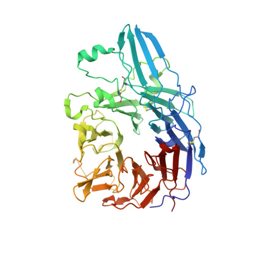
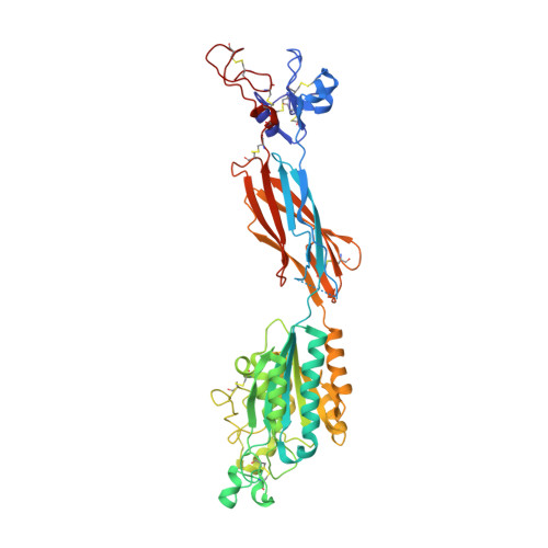
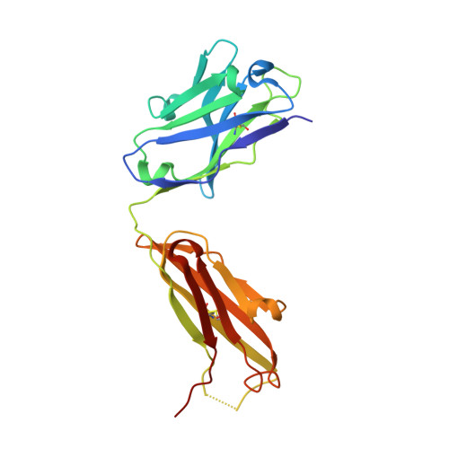
Structural Basis for Distinctive Recognition of Fibrinogen Gammac Peptide by the Platelet Integrin Alphaiibbeta3.

Springer, T.A.Zhu, J.Xiao, T.

(2008) J Cell Biol 182: 791

  • DOI: https://doi.org/10.1083/jcb.200801146
  • Primary Citation Related Structures: 
    2VC2, 2VDK, 2VDL, 2VDM, 2VDN, 2VDO, 2VDP, 2VDQ, 2VDR

  • PubMed Abstract: 

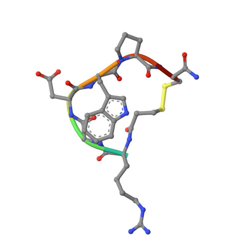
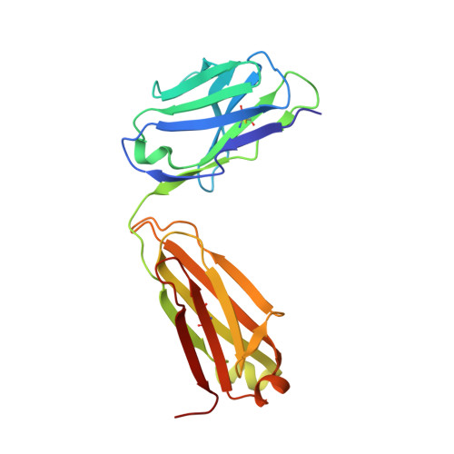
    Hemostasis and thrombosis (blood clotting) involve fibrinogen binding to integrin alpha(IIb)beta(3) on platelets, resulting in platelet aggregation. alpha(v)beta(3) binds fibrinogen via an Arg-Asp-Gly (RGD) motif in fibrinogen's alpha subunit. alpha(IIb)beta(3) also binds to fibrinogen; however, it does so via an unstructured RGD-lacking C-terminal region of the gamma subunit (gammaC peptide). These distinct modes of fibrinogen binding enable alpha(IIb)beta(3) and alpha(v)beta(3) to function cooperatively in hemostasis. In this study, crystal structures reveal the integrin alpha(IIb)beta(3)-gammaC peptide interface, and, for comparison, integrin alpha(IIb)beta(3) bound to a lamprey gammaC primordial RGD motif. Compared with RGD, the GAKQAGDV motif in gammaC adopts a different backbone configuration and binds over a more extended region. The integrin metal ion-dependent adhesion site (MIDAS) Mg(2+) ion binds the gammaC Asp side chain. The adjacent to MIDAS (ADMIDAS) Ca(2+) ion binds the gammaC C terminus, revealing a contribution for ADMIDAS in ligand binding. Structural data from this natively disordered gammaC peptide enhances our understanding of the involvement of gammaC peptide and integrin alpha(IIb)beta(3) in hemostasis and thrombosis.


  • Organizational Affiliation
    • Department of Pathology, Immune Disease Institute, Harvard Medical School, Boston, MA 02115, USA.

Macromolecule Content 

  • Total Structure Weight: 151.19 kDa 
  • Atom Count: 10,972 
  • Modeled Residue Count: 1,348 
  • Deposited Residue Count: 1,356 
  • Unique protein chains: 5

Macromolecules

Find similar proteins by:|  3D Structure
Entity ID: 1
MoleculeChains  Sequence LengthOrganismDetailsImage
INTEGRIN ALPHA-IIB452Homo sapiensMutation(s): 0 
UniProt & NIH Common Fund Data Resources
Find proteins for P08514 (Homo sapiens)
Explore P08514 
Go to UniProtKB:  P08514
PHAROS:  P08514
GTEx:  ENSG00000005961 
Entity Groups
Sequence Clusters30% Identity50% Identity70% Identity90% Identity95% Identity100% Identity
UniProt GroupP08514
Glycosylation
Glycosylation Sites: 2Go to GlyGen: P08514-1
Sequence Annotations
Expand
Reference Sequence
Find similar proteins by:|  3D Structure
Entity ID: 2
MoleculeChains  Sequence LengthOrganismDetailsImage
INTEGRIN BETA-3461Homo sapiensMutation(s): 0 
UniProt & NIH Common Fund Data Resources
Find proteins for P05106 (Homo sapiens)
Explore P05106 
Go to UniProtKB:  P05106
PHAROS:  P05106
GTEx:  ENSG00000259207 
Entity Groups
Sequence Clusters30% Identity50% Identity70% Identity90% Identity95% Identity100% Identity
UniProt GroupP05106
Glycosylation
Glycosylation Sites: 3Go to GlyGen: P05106-1
Sequence Annotations
Expand
Reference Sequence
Find similar proteins by:  Sequence   |   3D Structure  
Entity ID: 3
MoleculeChains  Sequence LengthOrganismDetailsImage
EPTIFIBATIDE8synthetic constructMutation(s): 0 
Find similar proteins by:|  3D Structure
Entity ID: 4
MoleculeChains  Sequence LengthOrganismDetailsImage
MONOCLONAL ANTIBODY 10E5 HEAVY CHAIND [auth H]221Mus musculusMutation(s): 0 
Find similar proteins by:|  3D Structure
Entity ID: 5
MoleculeChains  Sequence LengthOrganismDetailsImage
MONOCLONAL ANTIBODY 10E5 LIGHT CHAINE [auth L]214Mus musculusMutation(s): 0 
UniProt
Find proteins for P01837 (Mus musculus)
Explore P01837 
Go to UniProtKB:  P01837
Entity Groups
Sequence Clusters30% Identity50% Identity70% Identity90% Identity95% Identity100% Identity
UniProt GroupP01837
Sequence Annotations
Expand
Reference Sequence

Oligosaccharides

Help  
Entity ID: 6
MoleculeChains Length2D Diagram GlycosylationD Interactions
alpha-D-mannopyranose-(1-3)-[alpha-D-mannopyranose-(1-6)]alpha-D-mannopyranose-(1-4)-2-acetamido-2-deoxy-beta-D-glucopyranose-(1-4)-2-acetamido-2-deoxy-beta-D-glucopyranoseF [auth D]5N-Glycosylation
Glycosylation Resources
GlyTouCan: G21381MC
GlyCosmos: G21381MC
GlyGen: G21381MC
Entity ID: 7
MoleculeChains Length2D Diagram GlycosylationD Interactions
alpha-D-mannopyranose-(1-3)-[alpha-D-mannopyranose-(1-6)]alpha-D-mannopyranose-(1-6)-[alpha-D-mannopyranose-(1-3)]alpha-D-mannopyranose-(1-4)-2-acetamido-2-deoxy-beta-D-glucopyranose-(1-4)-2-acetamido-2-deoxy-beta-D-glucopyranoseG [auth E]7N-Glycosylation
Glycosylation Resources
GlyTouCan: G09609EN
GlyCosmos: G09609EN
GlyGen: G09609EN

Small Molecules

Ligands 4 Unique
IDChains Name / Formula / InChI Key2D Diagram3D Interactions
NAG

Query on NAG


Download:Ideal Coordinates CCD File
M [auth A],
N [auth A],
S [auth B]
2-acetamido-2-deoxy-beta-D-glucopyranose
C8 H15 N O6
OVRNDRQMDRJTHS-FMDGEEDCSA-N
GOL

Query on GOL


Download:Ideal Coordinates CCD File
H [auth A],
O [auth B]
GLYCEROL
C3 H8 O3
PEDCQBHIVMGVHV-UHFFFAOYSA-N
CA

Query on CA


Download:Ideal Coordinates CCD File
I [auth A]
J [auth A]
K [auth A]
L [auth A]
Q [auth B]
I [auth A],
J [auth A],
K [auth A],
L [auth A],
Q [auth B],
R [auth B]
CALCIUM ION
Ca
BHPQYMZQTOCNFJ-UHFFFAOYSA-N
MG

Query on MG


Download:Ideal Coordinates CCD File
P [auth B]MAGNESIUM ION
Mg
JLVVSXFLKOJNIY-UHFFFAOYSA-N
Modified Residues  1 Unique
IDChains TypeFormula2D DiagramParent
HRG
Query on HRG
C
L-PEPTIDE LINKINGC7 H16 N4 O2ARG

Experimental Data & Validation

Experimental Data

  • Method: X-RAY DIFFRACTION
  • Resolution: 2.90 Å
  • R-Value Free:  0.213 (Depositor), 0.216 (DCC) 
  • R-Value Work:  0.163 (Depositor), 0.170 (DCC) 
  • R-Value Observed: 0.166 (Depositor) 
Space Group: P 32 2 1
Unit Cell:
Length ( Å )Angle ( ˚ )
a = 149.593α = 90
b = 149.593β = 90
c = 175.683γ = 120
Software Package:
Software NamePurpose
REFMACrefinement
HKL-2000data reduction
SCALEPACKdata scaling
AMoREphasing

Structure Validation

View Full Validation Report


Ligand Structure Quality Assessment 


Entry History 

Deposition Data

Revision History  (Full details and data files)

  • Version 1.0: 2008-09-02
    Type: Initial release
  • Version 1.1: 2011-07-13
    Changes: Advisory, Version format compliance
  • Version 1.2: 2016-12-28
    Changes: Atomic model, Data collection, Database references, Derived calculations, Non-polymer description, Other, Source and taxonomy, Structure summary
  • Version 1.3: 2019-01-30
    Changes: Data collection, Experimental preparation, Other
  • Version 1.4: 2019-02-06
    Changes: Data collection, Experimental preparation
  • Version 1.5: 2019-11-20
    Changes: Advisory, Derived calculations, Other
  • Version 2.0: 2020-07-29
    Type: Remediation
    Reason: Carbohydrate remediation
    Changes: Advisory, Atomic model, Data collection, Derived calculations, Structure summary
  • Version 2.1: 2023-12-13
    Changes: Data collection, Database references, Derived calculations, Refinement description, Structure summary