2VAK

Crystal structure of the avian reovirus inner capsid protein sigmaA


Experimental Data Snapshot

  • Method: X-RAY DIFFRACTION
  • Resolution: 2.34 Å
  • R-Value Free: 0.271 
  • R-Value Work: 0.210 
  • R-Value Observed: 0.211 

wwPDB Validation   3D Report Full Report


This is version 1.5 of the entry. See complete history


Literature

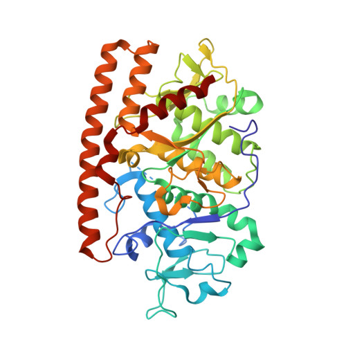
Crystal structure of the avian reovirus inner capsid protein sigmaA.

Guardado-Calvo, P.Vazquez-Iglesias, L.Martinez-Costas, J.Llamas-Saiz, A.L.Schoehn, G.Fox, G.C.Hermo-Parrado, X.L.Benavente, J.van Raaij, M.J.

(2008) J Virol 82: 11208-11216

  • DOI: https://doi.org/10.1128/JVI.00733-08
  • Primary Citation of Related Structures:  
    2VAK

  • PubMed Abstract: 

    Avian reovirus, an important avian pathogen, expresses eight structural and four nonstructural proteins. The structural sigmaA protein is a major component of the inner capsid, clamping together lambdaA building blocks. sigmaA has also been implicated in the resistance of avian reovirus to the antiviral action of interferon by strongly binding double-stranded RNA in the host cell cytoplasm and thus inhibiting activation of the double-stranded RNA-dependent protein kinase. We have solved the structure of bacterially expressed sigmaA by molecular replacement and refined it using data to 2.3-A resolution. Twelve sigmaA molecules are present in the P1 unit cell, arranged as two short double helical hexamers. A positively charged patch is apparent on the surface of sigmaA on the inside of this helix and mutation of either of two key arginine residues (Arg155 and Arg273) within this patch abolishes double-stranded RNA binding. The structural data, together with gel shift assay, electron microscopy, and sedimentation velocity centrifugation results, provide evidence for cooperative binding of sigmaA to double-stranded RNA. The minimal length of double-stranded RNA required for sigmaA binding was observed to be 14 to 18 bp.


  • Organizational Affiliation

    Departamento de Bioquímica e Bioloxía Molecular, Facultade de Farmacia, Universidade de Santiago de Compostela, E-15782 Santiago de Compostela, Spain.


Macromolecules
Find similar proteins by:  (by identity cutoff)  |  3D Structure
Entity ID: 1
MoleculeChains Sequence LengthOrganismDetailsImage
SIGMA A
A, B, C, D, E
A, B, C, D, E, F, G, H, I, J, K, L
423Avian orthoreovirusMutation(s): 0 
UniProt
Find proteins for Q9E6F8 (Avian orthoreovirus)
Explore Q9E6F8 
Go to UniProtKB:  Q9E6F8
Entity Groups  
Sequence Clusters30% Identity50% Identity70% Identity90% Identity95% Identity100% Identity
UniProt GroupQ9E6F8
Sequence Annotations
Expand
  • Reference Sequence
Small Molecules
Ligands 1 Unique
IDChains Name / Formula / InChI Key2D Diagram3D Interactions
SO4
Query on SO4

Download Ideal Coordinates CCD File 
M [auth A]
N [auth B]
O [auth C]
P [auth D]
Q [auth E]
M [auth A],
N [auth B],
O [auth C],
P [auth D],
Q [auth E],
R [auth F],
S [auth G],
T [auth H],
U [auth I],
V [auth J],
W [auth K],
X [auth L]
SULFATE ION
O4 S
QAOWNCQODCNURD-UHFFFAOYSA-L
Experimental Data & Validation

Experimental Data

  • Method: X-RAY DIFFRACTION
  • Resolution: 2.34 Å
  • R-Value Free: 0.271 
  • R-Value Work: 0.210 
  • R-Value Observed: 0.211 
  • Space Group: P 1
Unit Cell:
Length ( Å )Angle ( ˚ )
a = 103.21α = 93.81
b = 129.909β = 105.05
c = 144.037γ = 98.16
Software Package:
Software NamePurpose
REFMACrefinement
MOSFLMdata reduction
SCALAdata scaling
MOLREPphasing

Structure Validation

View Full Validation Report


Entry History 

Revision History  (Full details and data files)

  • Version 1.0: 2008-09-09
    Type: Initial release
  • Version 1.1: 2011-05-08
    Changes: Version format compliance
  • Version 1.2: 2011-07-13
    Changes: Version format compliance
  • Version 1.3: 2018-02-14
    Changes: Database references, Source and taxonomy
  • Version 1.4: 2019-05-08
    Changes: Data collection, Database references, Experimental preparation
  • Version 1.5: 2023-12-13
    Changes: Data collection, Database references, Derived calculations, Other, Refinement description