2J3Q | pdb_00002j3q

Torpedo acetylcholinesterase complexed with fluorophore thioflavin T


Experimental Data Snapshot

  • Method: X-RAY DIFFRACTION
  • Resolution: 2.80 Å
  • R-Value Free: 
    0.257 (Depositor), 0.248 (DCC) 
  • R-Value Work: 
    0.199 (Depositor), 0.192 (DCC) 
  • R-Value Observed: 
    0.199 (Depositor) 

Starting Model: experimental
View more details

wwPDB Validation 3D Report Full Report

Validation slider image for 2J3Q

Ligand Structure Quality Assessment 


This is version 2.3 of the entry. See complete history

Literature

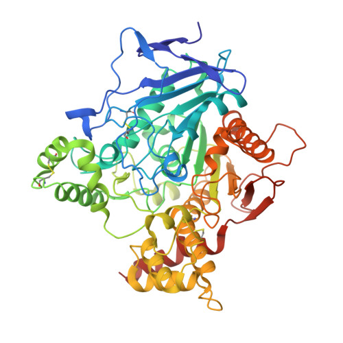
Crystal structure of thioflavin T bound to the peripheral site of Torpedo californica acetylcholinesterase reveals how thioflavin T acts as a sensitive fluorescent reporter of ligand binding to the acylation site.

Harel, M.Sonoda, L.K.Silman, I.Sussman, J.L.Rosenberry, T.L.

(2008) J Am Chem Soc 130: 7856-7861

  • DOI: https://doi.org/10.1021/ja7109822
  • Primary Citation Related Structures: 
    2J3Q

  • PubMed Abstract: 

    Acetylcholinesterase plays a key role in cholinergic synaptic transmission by hydrolyzing the neurotransmitter acetylcholine with one of the highest known catalytic rate constants. Hydrolysis occurs in a narrow and deep gorge that contains two sites of ligand binding: A peripheral site, or P-site, near the gorge entrance that contributes to catalytic efficiency both by transiently trapping substrate molecules as they enter the gorge and by allosterically accelerating the transfer of the substrate acyl group to a serine hydroxyl in an acylation site or A-site at the base of the gorge. Thioflavin T is a useful reporter of ligand interactions with the A-site. It binds specifically to the P-site with fluorescence that is enhanced approximately 1000-fold over that of unbound thioflavin T, and the enhanced fluorescence is quenched 1.5- to 4-fold when another ligand binds to the A-site in a ternary complex. To clarify the structural basis of this advantageous signal change, we here report the X-ray structure of the complex of thioflavin T with Torpedo californica acetylcholinesterase. The two aromatic rings in thioflavin T are coplanar and are packed snugly parallel to the aromatic side chains of Trp279, Tyr334, and Phe330. Overlays of this structure with the crystal structures of Torpedo californica acetylcholinesterase complexes with either edrophonium or m-( N, N, N-trimethylammonio)-2,2,2-trifluoroacetophenone, two small aromatic ligands that bind specifically to the A-site, indicate that the phenyl side chain of Phe330 must rotate to sterically accommodate both thioflavin T and the A-site ligand in the ternary complex. This rotation may allow some relaxation of the strict coplanarity of the aromatic rings in the bound thioflavin T and result in partial quenching of its fluorescence.


  • Organizational Affiliation
    • Department of Structural Biology, Weizmann Institute of Science, Rehovot 76100, Israel.

Macromolecule Content 

  • Total Structure Weight: 62.32 kDa 
  • Atom Count: 4,399 
  • Modeled Residue Count: 534 
  • Deposited Residue Count: 543 
  • Unique protein chains: 1

Macromolecules

Find similar proteins by:|  3D Structure
Entity ID: 1
MoleculeChains  Sequence LengthOrganismDetailsImage
ACETYLCHOLINESTERASE543Tetronarce californicaMutation(s): 0 
EC: 3.1.1.7
UniProt
Find proteins for P04058 (Tetronarce californica)
Explore P04058 
Go to UniProtKB:  P04058
Entity Groups
Sequence Clusters30% Identity50% Identity70% Identity90% Identity95% Identity100% Identity
UniProt GroupP04058
Glycosylation
Glycosylation Sites: 3
Sequence Annotations
Expand
Reference Sequence

Small Molecules

Ligands 3 Unique
IDChains Name / Formula / InChI Key2D Diagram3D Interactions
TFL

Query on TFL


Download:Ideal Coordinates CCD File
B [auth A]2-[4-(DIMETHYLAMINO)PHENYL]-6-HYDROXY-3-METHYL-1,3-BENZOTHIAZOL-3-IUM
C16 H17 N2 O S
NOVJJPLRUMZSDK-UHFFFAOYSA-O
NAG

Query on NAG


Download:Ideal Coordinates CCD File
C [auth A],
D [auth A],
E [auth A]
2-acetamido-2-deoxy-beta-D-glucopyranose
C8 H15 N O6
OVRNDRQMDRJTHS-FMDGEEDCSA-N
MG

Query on MG


Download:Ideal Coordinates CCD File
F [auth A],
G [auth A]
MAGNESIUM ION
Mg
JLVVSXFLKOJNIY-UHFFFAOYSA-N

Experimental Data & Validation

Experimental Data

  • Method: X-RAY DIFFRACTION
  • Resolution: 2.80 Å
  • R-Value Free:  0.257 (Depositor), 0.248 (DCC) 
  • R-Value Work:  0.199 (Depositor), 0.192 (DCC) 
  • R-Value Observed: 0.199 (Depositor) 
Space Group: P 32 2 1
Unit Cell:
Length ( Å )Angle ( ˚ )
a = 139.44α = 90
b = 139.44β = 90
c = 71.42γ = 120
Software Package:
Software NamePurpose
CNSrefinement
XDSdata reduction
XSCALEdata scaling
CNSphasing

Structure Validation

View Full Validation Report


Ligand Structure Quality Assessment 


Entry History 

Deposition Data

Revision History  (Full details and data files)

  • Version 1.0: 2007-09-04
    Type: Initial release
  • Version 1.1: 2011-05-08
    Changes: Version format compliance
  • Version 1.2: 2011-07-13
    Changes: Version format compliance
  • Version 2.0: 2018-07-25
    Changes: Advisory, Atomic model, Data collection, Database references, Derived calculations, Non-polymer description, Structure summary
  • Version 2.1: 2020-07-29
    Type: Remediation
    Reason: Carbohydrate remediation
    Changes: Data collection, Derived calculations, Other, Structure summary
  • Version 2.2: 2023-12-13
    Changes: Data collection, Database references, Refinement description, Structure summary
  • Version 2.3: 2024-10-16
    Changes: Structure summary