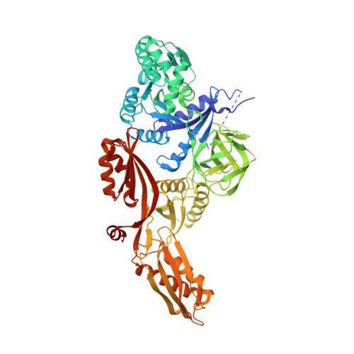
2BM0

Ribosomal elongation factor G (EF-G) Fusidic acid resistant mutant T84A


Experimental Data Snapshot

  • Method: X-RAY DIFFRACTION
  • Resolution: 2.40 Å
  • R-Value Free: 0.274 
  • R-Value Work: 0.210 
  • R-Value Observed: 0.210 

Starting Model: experimental
View more details

wwPDB Validation   3D Report Full Report


This is version 1.4 of the entry. See complete history


Literature

Structural Insights Into Fusidic Acid Resistance and Sensitivity in EF-G

Hansson, S.Singh, R.Gudkov, A.T.Liljas, A.Logan, D.T.

(2005) J Mol Biol 348: 939

  • DOI: https://doi.org/10.1016/j.jmb.2005.02.066
  • Primary Citation of Related Structures:  
    2BM0, 2BM1

  • PubMed Abstract: 

    Fusidic acid (FA) is a steroid antibiotic commonly used against Gram positive bacterial infections. It inhibits protein synthesis by stalling elongation factor G (EF-G) on the ribosome after translocation. A significant number of the mutations conferring strong FA resistance have been mapped at the interfaces between domains G, III and V of EF-G. However, direct information on how such mutations affect the structure has hitherto not been available. Here we present the crystal structures of two mutants of Thermus thermophilus EF-G, G16V and T84A, which exhibit FA hypersensitivity and resistance in vitro, respectively. These mutants also have higher and lower affinity for GTP respectively than wild-type EF-G. The mutations cause significant conformational changes in the switch II loop that have opposite effects on the position of a key residue, Phe90, which undergoes large conformational changes. This correlates with the importance of Phe90 in FA sensitivity reported in previous studies. These structures substantiate the importance of the domain G/domain III/domain V interfaces as a key component of the FA binding site. The mutations also cause subtle changes in the environment of the "P-loop lysine", Lys25. This led us to examine the conformation of the equivalent residue in all structures of translational GTPases, which revealed that EF-G and eEF2 form a group separate from the others and suggested that the role of Lys25 may be different in the two groups.


  • Organizational Affiliation

    Department of Molecular Biophysics, Lund University, Box 124, S-221 00 Lund, Sweden.


Macromolecules
Find similar proteins by:  (by identity cutoff)  |  3D Structure
Entity ID: 1
MoleculeChains Sequence LengthOrganismDetailsImage
ELONGATION FACTOR G691Thermus thermophilusMutation(s): 1 
UniProt
Find proteins for P13551 (Thermus thermophilus)
Explore P13551 
Go to UniProtKB:  P13551
Entity Groups  
Sequence Clusters30% Identity50% Identity70% Identity90% Identity95% Identity100% Identity
UniProt GroupP13551
Sequence Annotations
Expand
  • Reference Sequence
Small Molecules
Ligands 2 Unique
IDChains Name / Formula / InChI Key2D Diagram3D Interactions
GDP
Query on GDP

Download Ideal Coordinates CCD File 
B [auth A]GUANOSINE-5'-DIPHOSPHATE
C10 H15 N5 O11 P2
QGWNDRXFNXRZMB-UUOKFMHZSA-N
MG
Query on MG

Download Ideal Coordinates CCD File 
C [auth A]MAGNESIUM ION
Mg
JLVVSXFLKOJNIY-UHFFFAOYSA-N
Experimental Data & Validation

Experimental Data

  • Method: X-RAY DIFFRACTION
  • Resolution: 2.40 Å
  • R-Value Free: 0.274 
  • R-Value Work: 0.210 
  • R-Value Observed: 0.210 
  • Space Group: P 21 21 21
Unit Cell:
Length ( Å )Angle ( ˚ )
a = 78.2α = 90
b = 88.5β = 90
c = 116.9γ = 90
Software Package:
Software NamePurpose
XDSdata reduction
XDSdata scaling
MOLREPphasing
CCP4phasing
REFMACrefinement

Structure Validation

View Full Validation Report


Entry History 

Deposition Data

Revision History  (Full details and data files)

  • Version 1.0: 2005-05-04
    Type: Initial release
  • Version 1.1: 2011-05-08
    Changes: Version format compliance
  • Version 1.2: 2011-07-13
    Changes: Version format compliance
  • Version 1.3: 2018-01-17
    Changes: Data collection
  • Version 1.4: 2023-12-13
    Changes: Data collection, Database references, Derived calculations, Other, Refinement description