2BM1

Ribosomal elongation factor G (EF-G) Fusidic acid resistant mutant G16V


Experimental Data Snapshot

  • Method: X-RAY DIFFRACTION
  • Resolution: 2.60 Å
  • R-Value Free: 0.297 
  • R-Value Work: 0.220 
  • R-Value Observed: 0.220 

wwPDB Validation   3D Report Full Report


This is version 1.3 of the entry. See complete history


Literature

Structural Insights Into Fusidic Acid Resistance and Sensitivity in EF-G

Hansson, S.Singh, R.Gudkov, A.T.Liljas, A.Logan, D.T.

(2005) J Mol Biol 348: 939

  • DOI: https://doi.org/10.1016/j.jmb.2005.02.066
  • Primary Citation of Related Structures:  
    2BM0, 2BM1

  • PubMed Abstract: 

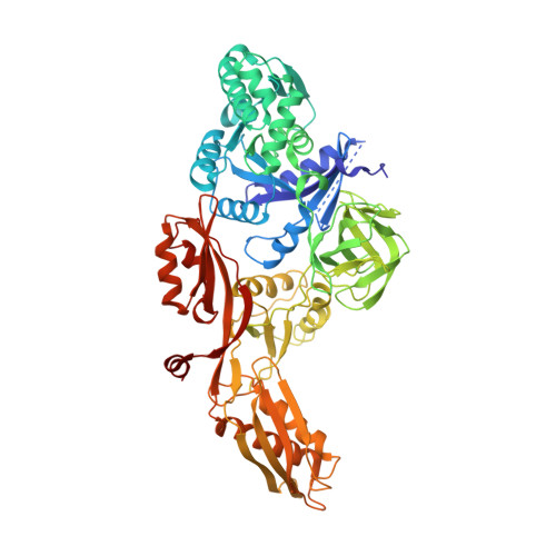
    Fusidic acid (FA) is a steroid antibiotic commonly used against Gram positive bacterial infections. It inhibits protein synthesis by stalling elongation factor G (EF-G) on the ribosome after translocation. A significant number of the mutations conferring strong FA resistance have been mapped at the interfaces between domains G, III and V of EF-G. However, direct information on how such mutations affect the structure has hitherto not been available. Here we present the crystal structures of two mutants of Thermus thermophilus EF-G, G16V and T84A, which exhibit FA hypersensitivity and resistance in vitro, respectively. These mutants also have higher and lower affinity for GTP respectively than wild-type EF-G. The mutations cause significant conformational changes in the switch II loop that have opposite effects on the position of a key residue, Phe90, which undergoes large conformational changes. This correlates with the importance of Phe90 in FA sensitivity reported in previous studies. These structures substantiate the importance of the domain G/domain III/domain V interfaces as a key component of the FA binding site. The mutations also cause subtle changes in the environment of the "P-loop lysine", Lys25. This led us to examine the conformation of the equivalent residue in all structures of translational GTPases, which revealed that EF-G and eEF2 form a group separate from the others and suggested that the role of Lys25 may be different in the two groups.


  • Organizational Affiliation

    Department of Molecular Biophysics, Lund University, Box 124, S-221 00 Lund, Sweden.


Macromolecules
Find similar proteins by:  (by identity cutoff)  |  3D Structure
Entity ID: 1
MoleculeChains Sequence LengthOrganismDetailsImage
ELONGATION FACTOR G691Thermus thermophilusMutation(s): 1 
UniProt
Find proteins for P13551 (Thermus thermophilus)
Explore P13551 
Go to UniProtKB:  P13551
Entity Groups  
Sequence Clusters30% Identity50% Identity70% Identity90% Identity95% Identity100% Identity
UniProt GroupP13551
Sequence Annotations
Expand
  • Reference Sequence
Small Molecules
Ligands 2 Unique
IDChains Name / Formula / InChI Key2D Diagram3D Interactions
GDP
Query on GDP

Download Ideal Coordinates CCD File 
B [auth A]GUANOSINE-5'-DIPHOSPHATE
C10 H15 N5 O11 P2
QGWNDRXFNXRZMB-UUOKFMHZSA-N
MG
Query on MG

Download Ideal Coordinates CCD File 
C [auth A]MAGNESIUM ION
Mg
JLVVSXFLKOJNIY-UHFFFAOYSA-N
Experimental Data & Validation

Experimental Data

  • Method: X-RAY DIFFRACTION
  • Resolution: 2.60 Å
  • R-Value Free: 0.297 
  • R-Value Work: 0.220 
  • R-Value Observed: 0.220 
  • Space Group: P 21 21 21
Unit Cell:
Length ( Å )Angle ( ˚ )
a = 76.3α = 90
b = 89.7β = 90
c = 114.9γ = 90
Software Package:
Software NamePurpose
REFMACrefinement
XDSdata reduction
SCALAdata scaling
MOLREPphasing

Structure Validation

View Full Validation Report


Entry History 

Deposition Data

Revision History  (Full details and data files)

  • Version 1.0: 2005-05-04
    Type: Initial release
  • Version 1.1: 2011-05-08
    Changes: Version format compliance
  • Version 1.2: 2011-07-13
    Changes: Version format compliance
  • Version 1.3: 2023-12-13
    Changes: Data collection, Database references, Derived calculations, Other, Refinement description