The importance of CH/pi hydrogen bonds in rational drug design: An ab initio fragment molecular orbital study to leukocyte-specific protein tyrosine (LCK) kinase
Ozawa, T., Tsuji, E., Ozawa, M., Handa, C., Mukaiyama, H., Nishimura, T., Kobayashi, S., Okazaki, K.(2008) Bioorg Med Chem 16: 10311-10318
- PubMed: 18977146 Search on PubMed
- DOI: https://doi.org/10.1016/j.bmc.2008.10.041
- Primary Citation Related Structures:
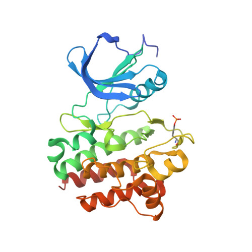
2ZM1, 2ZM4, 2ZYB - PubMed Abstract:
The interaction energy was calculated, by the ab initio FMO method, for complexes between LCK protein and four inhibitors (staurosporine, BMS compound 2, and our compounds 3 and 4). In every case a number of CH/pi hydrogen bonds have been disclosed in the so-called adenine pocket. In complexes of 2, 3, and 4, CH/pi and NH/pi hydrogen bonds have been observed in another pocket. In view of the above results, the aniline ring of 3 was replaced by 2,6-dimethyl aniline to increase the potency for LCK kinase. A 10-fold increase in the potency has been achieved for 4 over 3. We suggest that the concept of weak hydrogen bonds is useful in the rational design of drugs.
- Kissei Pharmaceutical Company Ltd., Central Research Laboratory, 4365-1 Kashiwabara, Hotaka, Azumino, Nagano-Pref 399-8304, Japan. tomonaga_ozawa@pharm.kissei.co.jp
Organizational Affiliation: