2Z1U | pdb_00002z1u

Crystal Structure of Hydrogenase Maturation Protein HypE in complex with ATP


Experimental Data Snapshot

  • Method: X-RAY DIFFRACTION
  • Resolution: 2.00 Å
  • R-Value Free: 
    0.228 (Depositor), 0.220 (DCC) 
  • R-Value Work: 
    0.200 (Depositor), 0.200 (DCC) 
  • R-Value Observed: 
    0.201 (Depositor) 

wwPDB Validation   3D Report Full Report


This is version 1.2 of the entry. See complete history


Literature

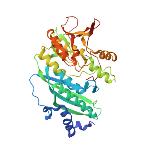
Crystal Structures of Hydrogenase Maturation Protein HypE in the Apo and ATP-bound Forms

Shomura, Y.Komori, H.Miyabe, N.Tomiyama, M.Shibata, N.Higuchi, Y.

(2007) J Mol Biology 372: 1045-1054

  • DOI: https://doi.org/10.1016/j.jmb.2007.07.023
  • Primary Citation of Related Structures:  
    2Z1T, 2Z1U

  • PubMed Abstract: 

    The hydrogenase maturation protein HypE serves an essential function in the biosynthesis of the nitrile group, which is subsequently coordinated to Fe as CN(-) ligands in [Ni-Fe] hydrogenase. Here, we present the crystal structures of HypE from Desulfovibrio vulgaris Hildenborough in the presence and in the absence of ATP at a resolution of 2.0 A and 2.6 A, respectively. Comparison of the apo structure with the ATP-bound structure reveals that binding ATP causes an induced-fit movement of the N-terminal portion, but does not entail an overall structural change. The residue Cys341 at the C terminus, whose thiol group is supposed to be carbamoylated before the nitrile group synthesis, is completely buried within the protein and is located in the vicinity of the gamma-phosphate group of the bound ATP. This suggests that the catalytic reaction occurs in this configuration but that a conformational change is required for the carbamoylation of Cys341. A glutamate residue is found close to the thiol group as well, which is suggestive of deprotonation of the carbamoyl group at the beginning of the reactions.


  • Organizational Affiliation

    Department of Life Science, Graduate School of Life Science, University of Hyogo, 3-2-1 Koto, Kamigori-cho, Ako-gun, Hyogo 678-1297, Japan; RIKEN SPring-8 Center, 1-1-1 Koto, Sayo-gun, Sayo-cho, Hyogo 679-5148, Japan. Electronic address: shomura@sci.u-hyogo.ac.jp.


Macromolecules
Find similar proteins by:  (by identity cutoff)  |  3D Structure
Entity ID: 1
MoleculeChains Sequence LengthOrganismDetailsImage
Hydrogenase expression/formation protein HypE343Nitratidesulfovibrio vulgaris str. HildenboroughMutation(s): 0 
Gene Names: hypE
UniProt
Find proteins for Q72F88 (Nitratidesulfovibrio vulgaris (strain ATCC 29579 / DSM 644 / CCUG 34227 / NCIMB 8303 / VKM B-1760 / Hildenborough))
Explore Q72F88 
Go to UniProtKB:  Q72F88
Entity Groups  
Sequence Clusters30% Identity50% Identity70% Identity90% Identity95% Identity100% Identity
UniProt GroupQ72F88
Sequence Annotations
Expand
  • Reference Sequence
Experimental Data & Validation

Experimental Data

  • Method: X-RAY DIFFRACTION
  • Resolution: 2.00 Å
  • R-Value Free:  0.228 (Depositor), 0.220 (DCC) 
  • R-Value Work:  0.200 (Depositor), 0.200 (DCC) 
  • R-Value Observed: 0.201 (Depositor) 
Space Group: P 31 2 1
Unit Cell:
Length ( Å )Angle ( ˚ )
a = 67.628α = 90
b = 67.628β = 90
c = 184.218γ = 120
Software Package:
Software NamePurpose
REFMACrefinement
HKL-2000data collection
HKL-2000data reduction
HKL-2000data scaling
MOLREPphasing

Structure Validation

View Full Validation Report


Entry History 

Deposition Data

Revision History  (Full details and data files)

  • Version 1.0: 2007-10-09
    Type: Initial release
  • Version 1.1: 2011-07-13
    Changes: Advisory, Derived calculations, Source and taxonomy, Version format compliance
  • Version 1.2: 2024-03-13
    Changes: Data collection, Database references, Derived calculations