Effect of a sodium ion on the dehydration-induced phase transition of monoclinic lysozyme crystals.
Harata, K., Akiba, T.(2007) Acta Crystallogr D Biol Crystallogr 63: 1016-1021
- PubMed: 17704571 Search on PubMed
- DOI: https://doi.org/10.1107/S0907444907031319
- Primary Citation Related Structures:
2Z12, 2Z18, 2Z19 - PubMed Abstract:
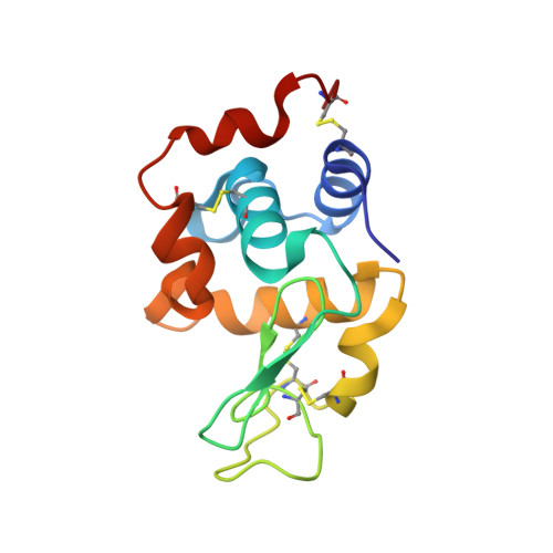
A monoclinic lysozyme crystal grown in NaCl solution was transformed into a new monoclinic crystal form by controlled dehydration. This crystal-to-crystal phase transition was accompanied by 20-40% solvent loss and the transformed crystal diffracted to prominently high resolution. The structures of the native and transformed crystals were determined at 1.4 and 1.15 A resolution, respectively. In the native crystal a sodium ion was bound to the loop region Ser60-Asn74; however, it was released in the transformed crystal and a water molecule occupied this position. In the transformed crystal a sodium ion was bound to the carboxyl group of Asp52, a catalytic residue. The same structural change was observed in the phase transition of a crystal soaked in a saturated NaCl solution. In contrast, a crystal soaked in 10% NaCl solution was transformed in a shorter time with a smaller loss of solvent and the structure of the sodium-binding site was conserved in the transformed crystal. The high concentration of NaCl is likely to stabilize the crystal structure against dehydration by forming salt linkages between protein molecules. This suggests that the sodium ion in the crystal regulates not only the structural change of the loop region Ser60-Asn74 but also the molecular rearrangement caused by dehydration.
- Biological Information Research Center, National Institute of Advanced Industrial Science and Technology (AIST), Central 6, 1-1-1 Higashi, Tsukuba, Ibaraki 305-8566, Japan. k-harata@aist.go.jp
Organizational Affiliation: