2YJE | pdb_00002yje

Oligomeric assembly of actin bound to MRTF-A


Experimental Data Snapshot

  • Method: X-RAY DIFFRACTION
  • Resolution: 3.10 Å
  • R-Value Free: 
    0.280 (Depositor), 0.283 (DCC) 
  • R-Value Work: 
    0.235 (Depositor), 0.236 (DCC) 
  • R-Value Observed: 
    0.237 (Depositor) 

Starting Model: experimental
View more details

wwPDB Validation 3D Report Full Report

Validation slider image for 2YJE

Ligand Structure Quality Assessment 


This is version 1.3 of the entry. See complete history

Literature

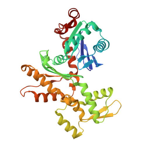
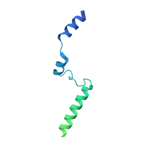
Structure of a pentavalent G-actin*MRTF-A complex reveals how G-actin controls nucleocytoplasmic shuttling of a transcriptional coactivator.

Mouilleron, S.Langer, C.A.Guettler, S.McDonald, N.Q.Treisman, R.

(2011) Sci Signal 4: ra40-ra40

  • DOI: https://doi.org/10.1126/scisignal.2001750
  • Primary Citation Related Structures: 
    2YJE, 2YJF

  • PubMed Abstract: 

    Subcellular localization of the actin-binding transcriptional coactivator MRTF-A is controlled by its interaction with monomeric actin (G-actin). Signal-induced decreases in G-actin concentration reduce MRTF-A nuclear export, leading to its nuclear accumulation, whereas artificial increases in G-actin concentration in resting cells block MRTF-A nuclear import, retaining it in the cytoplasm. This regulation is dependent on three actin-binding RPEL motifs in the regulatory domain of MRTF-A. We describe the structures of pentavalent and trivalent G-actin•RPEL domain complexes. In the pentavalent complex, each RPEL motif and the two intervening spacer sequences bound an actin monomer, forming a compact assembly. In contrast, the trivalent complex lacked the C-terminal spacer- and RPEL-actins, both of which bound only weakly in the pentavalent complex. Cytoplasmic localization of MRTF-A in unstimulated fibroblasts also required binding of G-actin to the spacer sequences. The bipartite MRTF-A nuclear localization sequence was buried in the pentameric assembly, explaining how increases in G-actin concentration prevent nuclear import of MRTF-A. Analyses of the pentavalent and trivalent complexes show how actin loads onto the RPEL domain and reveal a molecular mechanism by which actin can control the activity of one of its binding partners.


  • Organizational Affiliation
    • Structural Biology Group, Cancer Research UK London Research Institute, Lincoln's Inn Fields Laboratories, 44 Lincoln's Inn Fields, London WC2A 3LY, UK.

Macromolecule Content 

  • Total Structure Weight: 144.85 kDa 
  • Atom Count: 8,199 
  • Modeled Residue Count: 1,081 
  • Deposited Residue Count: 1,268 
  • Unique protein chains: 2

Macromolecules

Find similar proteins by:|  3D Structure
Entity ID: 1
MoleculeChains  Sequence LengthOrganismDetailsImage
ACTIN, ALPHA SKELETAL MUSCLE
A, B, C
377Oryctolagus cuniculusMutation(s): 0 
EC: 3.6.4
UniProt
Find proteins for P68135 (Oryctolagus cuniculus)
Explore P68135 
Go to UniProtKB:  P68135
Entity Groups
Sequence Clusters30% Identity50% Identity70% Identity90% Identity95% Identity100% Identity
UniProt GroupP68135
Sequence Annotations
Expand
Reference Sequence
Find similar proteins by:|  3D Structure
Entity ID: 2
MoleculeChains  Sequence LengthOrganismDetailsImage
MKL/MYOCARDIN-LIKE PROTEIN 1D [auth M]137Mus musculusMutation(s): 0 
UniProt
Find proteins for Q8K4J6 (Mus musculus)
Explore Q8K4J6 
Go to UniProtKB:  Q8K4J6
Entity Groups
Sequence Clusters30% Identity50% Identity70% Identity90% Identity95% Identity100% Identity
UniProt GroupQ8K4J6
Sequence Annotations
Expand
Reference Sequence

Small Molecules

Ligands 3 Unique
IDChains Name / Formula / InChI Key2D Diagram3D Interactions
ATP

Query on ATP


Download:Ideal Coordinates CCD File
F [auth A],
I [auth B],
L [auth C]
ADENOSINE-5'-TRIPHOSPHATE
C10 H16 N5 O13 P3
ZKHQWZAMYRWXGA-KQYNXXCUSA-N
LAB

Query on LAB


Download:Ideal Coordinates CCD File
E [auth A],
H [auth B],
K [auth C]
LATRUNCULIN B
C20 H29 N O5 S
NSHPHXHGRHSMIK-JRIKCGFMSA-N
MG

Query on MG


Download:Ideal Coordinates CCD File
G [auth A],
J [auth B],
M [auth C]
MAGNESIUM ION
Mg
JLVVSXFLKOJNIY-UHFFFAOYSA-N

Experimental Data & Validation

Experimental Data

  • Method: X-RAY DIFFRACTION
  • Resolution: 3.10 Å
  • R-Value Free:  0.280 (Depositor), 0.283 (DCC) 
  • R-Value Work:  0.235 (Depositor), 0.236 (DCC) 
  • R-Value Observed: 0.237 (Depositor) 
Space Group: P 43 21 2
Unit Cell:
Length ( Å )Angle ( ˚ )
a = 90.935α = 90
b = 90.935β = 90
c = 321.613γ = 90
Software Package:
Software NamePurpose
PHENIXrefinement
PHENIXphasing

Structure Validation

View Full Validation Report


Ligand Structure Quality Assessment 


Entry History 

Deposition Data

Revision History  (Full details and data files)

  • Version 1.0: 2011-07-06
    Type: Initial release
  • Version 1.1: 2011-11-16
    Changes: Database references, Other, Version format compliance
  • Version 1.2: 2018-04-11
    Changes: Data collection, Database references
  • Version 1.3: 2023-12-20
    Changes: Data collection, Database references, Derived calculations, Other, Refinement description