Raman Assisted Crystallography Reveals a Mechanism of X-Ray Induced Reversible Disulfide Radical Formation
Carpentier, P., Royant, A., Weik, M., Bourgeois, D.(2010) Structure 18: 1410
- PubMed: 21070940 Search on PubMed
- DOI: https://doi.org/10.1016/j.str.2010.09.010
- Primary Citation Related Structures:
2XBR, 2XBS - PubMed Abstract:
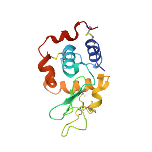
X-ray-induced chemistry modifies biological macromolecules structurally and functionally, even at cryotemperatures. The mechanisms of x-radiation damage in colored or redox proteins have often been investigated by combining X-ray crystallography with in crystallo-ultraviolet-visible spectroscopy. Here, we used Raman microspectrophotometry to follow the onset of damage in crystalline lysozyme, notably that of disulfide bond breakage. The dose-dependent Raman spectra are consistent with a kinetic model for the rupture of disulfide bonds suggesting the rapid build up of an anionic radical intermediate. This intermediate may either revert back to the oxidized state or evolve toward protonated radical species or cleaved products. The data strongly suggest that back conversion of the anionic radical is significantly accelerated by X-rays, revealing an X-ray-induced "repair" mechanism. The possibility of X-ray-induced chemical repair is an important feature to take into account when assessing radiation damage in macromolecules.
- Laboratoire de Cristallogenèse et Cristallographie des Protéines, IBS, Institut de Biologie Structurale Jean-Pierre Ebel, CEA, CNRS, Université Joseph Fourier, 41 Rue Jules Horowitz, F-38027 Grenoble, France. philippe.carpentier@ibs.fr
Organizational Affiliation: