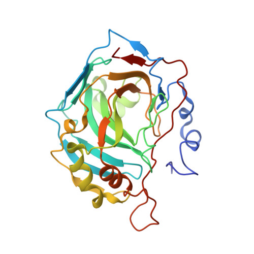
Structures of Human Carbonic Anhydrase II/Inhibitor Complexes Reveal a Second Binding Site for Steroidal and Non-Steroidal Inhibitors.
Cozier, G.E., Leese, M.P., Lloyd, M.D., Baker, M.D., Thiyagarajan, N., Acharya, K.R., Potter, B.V.L.(2010) Biochemistry 49: 3464
- PubMed: 20297840
- DOI: https://doi.org/10.1021/bi902178w
- Primary Citation of Related Structures:
2X7S, 2X7T, 2X7U - PubMed Abstract:
Carbonic anhydrase (CA) catalyzes the reversible hydration of carbon dioxide to hydrogen carbonate, and its role in maintaining pH balance has made it an attractive drug target. Steroidal sulfamate esters, inhibitors of the cancer drug target steroid sulfatase (STS), are sequestered in vivo by CA II in red blood cells, which may be the origin of their excellent drug properties. Understanding the structural basis of this is important for drug design. Structures of CA II complexed with 2-methoxyestradiol 3-O-sulfamate (3), 2-ethylestradiol 3,17-O,O-bis(sulfamate) (4), and 2-methoxyestradiol 17-O-sulfamate (5) are reported to 2.10, 1.85, and 1.64 A, respectively. Inhibitor 3 interacts with the active site Zn(II) ion through the 3-O-sulfamate, while inhibitors 4 and 5 bind through their 17-O-sulfamate. Comparison of the IC(50) values for CA II inhibition gave respective values of 56, 662, 2113, 169, 770, and 86 nM for estrone 3-O-sulfamate (1), 2-methoxyestradiol 3,17-O,O-bis(sulfamate) (2), 3, 4, 5, and 5'-((4H-1,2,4-triazol-4-yl)methyl)-3-chloro-2'-cyanobiphenyl-4-yl sulfamate (6), a nonsteroidal dual aromatase-sulfatase inhibitor. Inhibitors 2, 5, and 6 showed binding to a second adjacent site that is capable of binding both steroidal and nonsteroidal ligands. Examination of both IC(50) values and crystal structures suggests that 2-substituents on the steroid nucleus hinder binding via a 3-O-sulfamate, leading to coordination through a 17-O-sulfamate if present. These results underline the influence of small structural changes on affinity and mode of binding, the degree of flexibility in the design of sulfamate-based inhibitors, and suggest a strategy for inhibitors which interact with both the active site and the second adjacent binding site simultaneously that could be both potent and selective.
- Medicinal Chemistry, Department of Pharmacy and Pharmacology, University of Bath,Claverton Down, Bath BA2 7AY, United Kingdom.
Organizational Affiliation:


