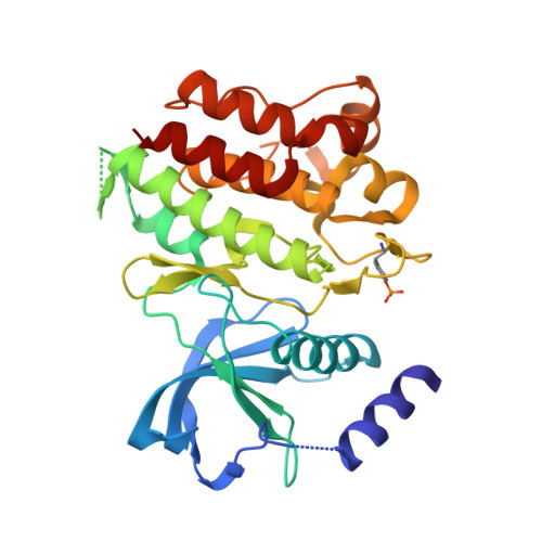
2X2L

Crystal Structure of phosphorylated RET tyrosine kinase domain with inhibitor


Experimental Data Snapshot

  • Method: X-RAY DIFFRACTION
  • Resolution: 2.00 Å
  • R-Value Free: 0.269 
  • R-Value Work: 0.211 
  • R-Value Observed: 0.214 

Starting Model: experimental
View more details

wwPDB Validation   3D Report Full Report


Ligand Structure Quality Assessment 


This is version 1.6 of the entry. See complete history


Literature

Synthesis, structure-activity relationship and crystallographic studies of 3-substituted indolin-2-one RET inhibitors.

Mologni, L.Rostagno, R.Brussolo, S.Knowles, P.P.Kjaer, S.Murray-Rust, J.Rosso, E.Zambon, A.Scapozza, L.McDonald, N.Q.Lucchini, V.Gambacorti-Passerini, C.

(2010) Bioorg Med Chem 18: 1482-1496

  • DOI: https://doi.org/10.1016/j.bmc.2010.01.011
  • Primary Citation of Related Structures:  
    2X2K, 2X2L, 2X2M

  • PubMed Abstract: 

    The synthesis, structure-activity relationships (SAR) and structural data of a series of indolin-2-one inhibitors of RET tyrosine kinase are described. These compounds were designed to explore the available space around the indolinone scaffold within RET active site. Several substitutions at different positions were tested and biochemical data were used to draw a molecular model of steric and electrostatic interactions, which can be applied to design more potent and selective RET inhibitors. The crystal structures of RET kinase domain in complex with three inhibitors were solved. All three compounds bound in the ATP pocket and formed two hydrogen bonds with the kinase hinge region. Crystallographic analysis confirmed predictions from molecular modelling and helped refine SAR results. These data provide important information for the development of indolinone inhibitors for the treatment of RET-driven cancers.


  • Organizational Affiliation

    Department of Clinical Medicine and Prevention, University of Milano-Bicocca, Monza, Italy. luca.mologni@unimib.it


Macromolecules
Find similar proteins by:  (by identity cutoff)  |  3D Structure
Entity ID: 1
MoleculeChains Sequence LengthOrganismDetailsImage
PROTO-ONCOGENE TYROSINE-PROTEIN KINASE RECEPTOR RET314Homo sapiensMutation(s): 0 
EC: 2.7.10.1
UniProt & NIH Common Fund Data Resources
Find proteins for P07949 (Homo sapiens)
Explore P07949 
Go to UniProtKB:  P07949
PHAROS:  P07949
GTEx:  ENSG00000165731 
Entity Groups  
Sequence Clusters30% Identity50% Identity70% Identity90% Identity95% Identity100% Identity
UniProt GroupP07949
Sequence Annotations
Expand
  • Reference Sequence
Small Molecules
Modified Residues  1 Unique
IDChains TypeFormula2D DiagramParent
PTR
Query on PTR
A
L-PEPTIDE LINKINGC9 H12 N O6 PTYR
Binding Affinity Annotations 
IDSourceBinding Affinity
X2L BindingDB:  2X2L IC50: min: 5300, max: 6.30e+4 (nM) from 2 assay(s)
PDBBind:  2X2L IC50: 5300 (nM) from 1 assay(s)
Experimental Data & Validation

Experimental Data

  • Method: X-RAY DIFFRACTION
  • Resolution: 2.00 Å
  • R-Value Free: 0.269 
  • R-Value Work: 0.211 
  • R-Value Observed: 0.214 
  • Space Group: C 1 2 1
Unit Cell:
Length ( Å )Angle ( ˚ )
a = 71.999α = 90
b = 70.685β = 110.35
c = 71.573γ = 90
Software Package:
Software NamePurpose
REFMACrefinement
MOSFLMdata reduction
SCALAdata scaling
MOLREPphasing

Structure Validation

View Full Validation Report


Ligand Structure Quality Assessment 


Entry History 

Deposition Data

Revision History  (Full details and data files)

  • Version 1.0: 2010-02-09
    Type: Initial release
  • Version 1.1: 2011-05-07
    Changes: Version format compliance
  • Version 1.2: 2011-07-13
    Changes: Version format compliance
  • Version 1.3: 2018-02-28
    Changes: Database references
  • Version 1.4: 2019-01-30
    Changes: Data collection, Experimental preparation
  • Version 1.5: 2019-05-08
    Changes: Data collection, Derived calculations, Experimental preparation
  • Version 1.6: 2023-12-20
    Changes: Data collection, Database references, Derived calculations, Other, Refinement description