Crystal Structure of a Class Xib Phospholipase A2 (Pla2): Rice (Oryza Sativa) Isoform-2 Pla2 and an Octanoate Complex.
Guy, J.E., Stahl, U., Lindqvist, Y.(2009) J Biol Chem 284: 19371
- PubMed: 19457861 Search on PubMedSearch on PubMed Central
- DOI: https://doi.org/10.1074/jbc.M109.008466
- Primary Citation Related Structures:
2WG7, 2WG8, 2WG9 - PubMed Abstract:
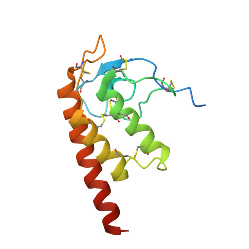
Phospholipase A(2) catalyzes the specific hydrolysis of the sn-2 acyl bond of various glycerophospholipids, producing fatty acids and lysophospholipids. Phospholipase A(2)s (PLA(2)s) constitute a large superfamily of enzymes whose products are important for a multitude of signal transduction processes, lipid mediator release, lipid metabolism, development, plant stress responses, and host defense. The crystal structure of rice (Oryza sativa) isoform 2 phospholipase A(2) has been determined to 2.0 A resolution using sulfur SAD phasing, and shows that the class XIb phospholipases have a unique structure compared with other secreted PLA(2)s. The N-terminal half of the chain contains mainly loop structure, including the conserved Ca(2+)-binding loop, but starts with a short 3(10)-helix and also includes two short anti-parallel beta-strands. The C-terminal half is folded into three anti-parallel alpha-helices, of which the two first are also present in other secreted PLA(2)s and contain the conserved catalytic histidine and calcium liganding aspartate residues. The structure is stabilized by six disulfide bonds. The water structure around the calcium ion binding site suggests the involvement of a second water molecule in the mechanism for hydrolysis, the water-assisted calcium-coordinate oxyanion mechanism. The octanoate molecule in the complex structure is bound in a hydrophobic pocket, which extends to the likely membrane interface and is proposed to model the binding of the product fatty acid. Due to the differences in structure, the suggested surface for binding to the membrane has a different morphology in the rice PLA(2) compared with other phospholipases.
- Department of Medical Biochemistry and Biophysics, Karolinska Institutet, Stockholm S-17177, Sweden.
Organizational Affiliation: