2W2X

Complex of Rac2 and PLCg2 spPH Domain


Experimental Data Snapshot

  • Method: X-RAY DIFFRACTION
  • Resolution: 2.30 Å
  • R-Value Free: 0.294 
  • R-Value Work: 0.234 
  • R-Value Observed: 0.237 

wwPDB Validation   3D Report Full Report


Ligand Structure Quality Assessment 


This is version 1.3 of the entry. See complete history


Literature

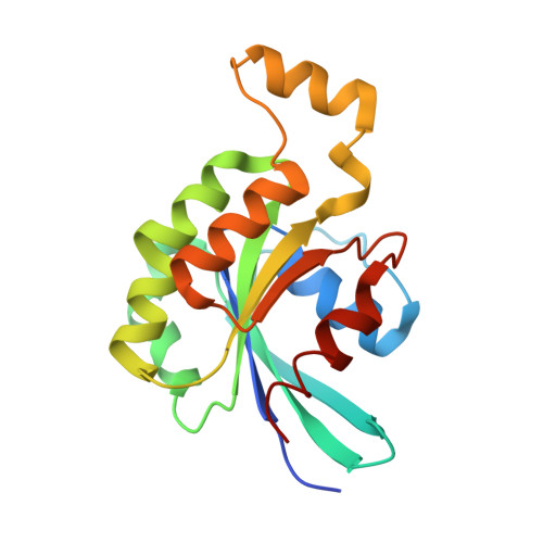
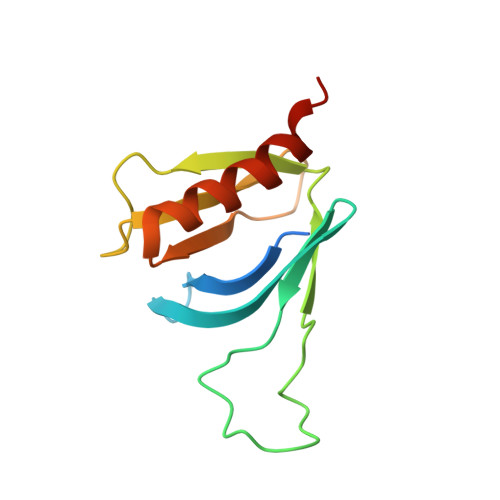
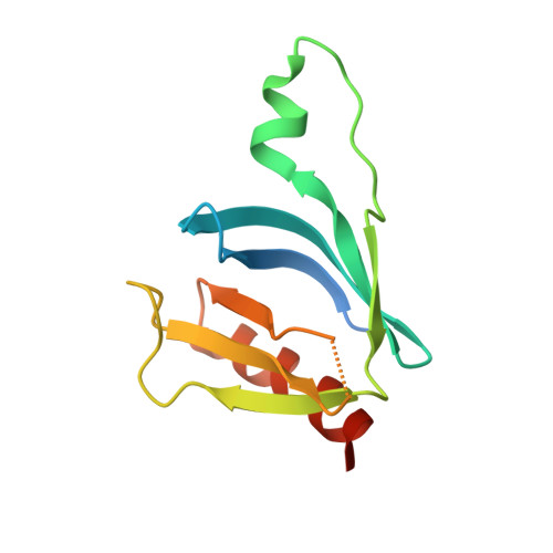
Structural Insights Into Formation of an Active Signaling Complex between Rac and Phospholipase C Gamma 2.

Bunney, T.D.Opaleye, O.Roe, S.M.Vatter, P.Baxendale, R.W.Walliser, C.Everett, K.L.Josephs, M.B.Christow, C.Rodrigues-Lima, F.Gierschik, P.Pearl, L.H.Katan, M.

(2009) Mol Cell 34: 223

  • DOI: https://doi.org/10.1016/j.molcel.2009.02.023
  • Primary Citation of Related Structures:  
    2W2T, 2W2V, 2W2W, 2W2X

  • PubMed Abstract: 
  • Rho family GTPases are important cellular switches and control a number of physiological functions. Understanding the molecular basis of interaction of these GTPases with their effectors is crucial in understanding their functions in the cell. Here we present the crystal structure of the complex of Rac2 bound to the split pleckstrin homology (spPH) domain of phospholipase C-gamma(2) (PLCgamma(2)) ...

    Rho family GTPases are important cellular switches and control a number of physiological functions. Understanding the molecular basis of interaction of these GTPases with their effectors is crucial in understanding their functions in the cell. Here we present the crystal structure of the complex of Rac2 bound to the split pleckstrin homology (spPH) domain of phospholipase C-gamma(2) (PLCgamma(2)). Based on this structure, we illustrate distinct requirements for PLCgamma(2) activation by Rac and EGF and generate Rac effector mutants that specifically block activation of PLCgamma(2), but not the related PLCbeta(2) isoform. Furthermore, in addition to the complex, we report the crystal structures of free spPH and Rac2 bound to GDP and GTPgammaS. These structures illustrate a mechanism of conformational switches that accompany formation of signaling active complexes and highlight the role of effector binding as a common feature of Rac and Cdc42 interactions with a variety of effectors.


    Organizational Affiliation

    Section of Cell and Molecular Biology , The Institute of Cancer Research, London, UK. tom.bunney@icr.ac.uk


Macromolecules
Find similar proteins by:  (by identity cutoff)  |  3D Structure
Entity ID: 1
MoleculeChainsSequence LengthOrganismDetailsImage
RAS-RELATED C3 BOTULINUM TOXIN SUBSTRATE 2
A, B
185Homo sapiensMutation(s): 1 
Gene Names: RAC2
UniProt & NIH Common Fund Data Resources
Find proteins for P15153 (Homo sapiens)
Explore P15153 
Go to UniProtKB:  P15153
PHAROS:  P15153
Entity Groups  
Sequence Clusters30% Identity50% Identity70% Identity90% Identity95% Identity100% Identity
UniProt GroupP15153
Protein Feature View
Expand
  • Reference Sequence
Find similar proteins by:  (by identity cutoff)  |  3D Structure
Entity ID: 2
MoleculeChainsSequence LengthOrganismDetailsImage
1-PHOSPHATIDYLINOSITOL-4,5-BISPHOSPHATE PHOSPHODIESTERASE GAMMA-2124Homo sapiensMutation(s): 0 
Gene Names: PLCG2
EC: 3.1.4.11
UniProt & NIH Common Fund Data Resources
Find proteins for P16885 (Homo sapiens)
Explore P16885 
Go to UniProtKB:  P16885
PHAROS:  P16885
Entity Groups  
Sequence Clusters30% Identity50% Identity70% Identity90% Identity95% Identity100% Identity
UniProt GroupP16885
Protein Feature View
Expand
  • Reference Sequence
Find similar proteins by:  (by identity cutoff)  |  3D Structure
Entity ID: 3
MoleculeChainsSequence LengthOrganismDetailsImage
1-PHOSPHATIDYLINOSITOL-4,5-BISPHOSPHATE PHOSPHODIESTERASE GAMMA-2124Homo sapiensMutation(s): 0 
Gene Names: PLCG2
EC: 3.1.4.11
UniProt & NIH Common Fund Data Resources
Find proteins for P16885 (Homo sapiens)
Explore P16885 
Go to UniProtKB:  P16885
PHAROS:  P16885
Entity Groups  
Sequence Clusters30% Identity50% Identity70% Identity90% Identity95% Identity100% Identity
UniProt GroupP16885
Protein Feature View
Expand
  • Reference Sequence
Small Molecules
Ligands 2 Unique
IDChainsName / Formula / InChI Key2D Diagram3D Interactions
GSP
Query on GSP

Download Ideal Coordinates CCD File 
E [auth A],
G [auth B]
5'-GUANOSINE-DIPHOSPHATE-MONOTHIOPHOSPHATE
C10 H16 N5 O13 P3 S
XOFLBQFBSOEHOG-UUOKFMHZSA-N
 Ligand Interaction
MG
Query on MG

Download Ideal Coordinates CCD File 
F [auth A],
H [auth B]
MAGNESIUM ION
Mg
JLVVSXFLKOJNIY-UHFFFAOYSA-N
 Ligand Interaction
Experimental Data & Validation

Experimental Data

  • Method: X-RAY DIFFRACTION
  • Resolution: 2.30 Å
  • R-Value Free: 0.294 
  • R-Value Work: 0.234 
  • R-Value Observed: 0.237 
  • Space Group: C 1 2 1
Unit Cell:
Length ( Å )Angle ( ˚ )
a = 131.57α = 90
b = 84.459β = 112.21
c = 74.196γ = 90
Software Package:
Software NamePurpose
PHENIXrefinement
MOSFLMdata reduction
SCALAdata scaling
PHASERphasing

Structure Validation

View Full Validation Report


Ligand Structure Quality Assessment 


Entry History 

Deposition Data

Revision History  (Full details and data files)

  • Version 1.0: 2009-05-05
    Type: Initial release
  • Version 1.1: 2011-05-08
    Changes: Version format compliance
  • Version 1.2: 2011-07-13
    Changes: Version format compliance
  • Version 1.3: 2019-05-15
    Changes: Data collection, Experimental preparation, Other