2VH9

CRYSTAL STRUCTURE OF NXG1-DELTAYNIIG IN COMPLEX WITH XLLG, A XYLOGLUCAN DERIVED OLIGOSACCHARIDE


Experimental Data Snapshot

  • Method: X-RAY DIFFRACTION
  • Resolution: 2.10 Å
  • R-Value Free: 0.203 
  • R-Value Work: 0.148 
  • R-Value Observed: 0.151 

wwPDB Validation   3D Report Full Report


Ligand Structure Quality Assessment 


This is version 2.1 of the entry. See complete history


Literature

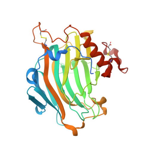
Analysis of Nasturtium Tmnxg1 Complexes by Crystallography and Molecular Dynamics Provides Detailed Insight Into Substrate Recognition by Family Gh16 Xyloglucan Endo-Transglycosylases and Endo-Hydrolases.

Mark, P.Baumann, M.J.Eklof, J.M.Gullfot, F.Michel, G.Kallas, A.M.Teeri, T.T.Brumer, H.Czjzek, M.

(2009) Proteins 75: 820

  • DOI: https://doi.org/10.1002/prot.22291
  • Primary Citation of Related Structures:  
    2VH9

  • PubMed Abstract: 

    Reorganization and degradation of the wall crosslinking and seed storage polysaccharide xyloglucan by glycoside hydrolase family 16 (GH16) endo-transglycosylases and hydrolases are crucial to the growth of the majority of land plants, affecting processes as diverse as germination, morphogenesis, and fruit ripening. A high-resolution, three-dimensional structure of a nasturtium (Tropaeolum majus) endo-xyloglucanase loop mutant, TmNXG1-DeltaYNIIG, with an oligosaccharide product bound in the negative active-site subsites, has been solved by X-ray crystallography. Comparison of this novel complex to that of the strict xyloglucan endo-transglycosylase PttXET16-34 from hybrid aspen (Populus tremula x tremuloides), previously solved with a xylogluco-oligosaccharide bound in the positive subsites, highlighted key protein structures that affect the disparate catalytic activities displayed by these closely related enzymes. Combination of these "partial" active-site complexes through molecular dynamics simulations in water allowed modeling of wild-type TmNXG1, TmNXG1-DeltaYNIIG, and wild-type PttXET16-34 in complex with a xyloglucan octadecasaccharide spanning the entire catalytic cleft. A comprehensive analysis of these full-length complexes underscored the importance of various loops lining the active site. Subtle differences leading to a tighter hydrogen bonding pattern on the negative (glycosyl donor) binding subsites, together with loop flexibility on the positive (glycosyl acceptor) binding subsites appear to favor hydrolysis over transglycosylation in GH16 xyloglucan-active enzymes.


  • Organizational Affiliation

    School of Biotechnology, Royal Institute of Technology (KTH), AlbaNova University Centre. S 106 91 Stockholm, Sweden.


Macromolecules
Find similar proteins by:  (by identity cutoff)  |  3D Structure
Entity ID: 1
MoleculeChains Sequence LengthOrganismDetailsImage
CELLULASE
A, B
290Tropaeolum majusMutation(s): 0 
EC: 3.2.1.151 (PDB Primary Data), 3.2.1.4 (PDB Primary Data)
UniProt
Find proteins for Q07524 (Tropaeolum majus)
Explore Q07524 
Go to UniProtKB:  Q07524
Entity Groups  
Sequence Clusters30% Identity50% Identity70% Identity90% Identity95% Identity100% Identity
UniProt GroupQ07524
Sequence Annotations
Expand
  • Reference Sequence
Oligosaccharides

Help

Entity ID: 2
MoleculeChains Length2D Diagram Glycosylation3D Interactions
beta-D-galactopyranose-(1-2)-alpha-D-xylopyranose-(1-6)-beta-D-glucopyranose
C, D, F, G
3N/A
Glycosylation Resources
GlyTouCan:  G95959IY
GlyCosmos:  G95959IY
Entity ID: 3
MoleculeChains Length2D Diagram Glycosylation3D Interactions
alpha-D-xylopyranose-(1-6)-beta-D-glucopyranose
E, H
2N/A
Glycosylation Resources
GlyTouCan:  G00977CB
GlyCosmos:  G00977CB
GlyGen:  G00977CB
Experimental Data & Validation

Experimental Data

  • Method: X-RAY DIFFRACTION
  • Resolution: 2.10 Å
  • R-Value Free: 0.203 
  • R-Value Work: 0.148 
  • R-Value Observed: 0.151 
  • Space Group: P 31
Unit Cell:
Length ( Å )Angle ( ˚ )
a = 100.54α = 90
b = 100.54β = 90
c = 61.13γ = 120
Software Package:
Software NamePurpose
REFMACrefinement
MOSFLMdata reduction
SCALAdata scaling
AMoREphasing

Structure Validation

View Full Validation Report


Ligand Structure Quality Assessment 


Entry History 

Deposition Data

Revision History  (Full details and data files)

  • Version 1.0: 2008-11-25
    Type: Initial release
  • Version 1.1: 2011-08-17
    Changes: Atomic model, Derived calculations, Non-polymer description, Structure summary, Version format compliance
  • Version 1.2: 2017-07-12
    Changes: Derived calculations
  • Version 1.3: 2019-05-08
    Changes: Data collection, Experimental preparation
  • Version 2.0: 2020-07-29
    Type: Remediation
    Reason: Carbohydrate remediation
    Changes: Advisory, Atomic model, Data collection, Derived calculations, Other, Structure summary
  • Version 2.1: 2023-12-13
    Changes: Data collection, Database references, Derived calculations, Refinement description, Structure summary