2Q7T

Crystal Structure of the F plasmid TraI Relaxase Domain with the Scissile Thymidine Base


Experimental Data Snapshot

  • Method: X-RAY DIFFRACTION
  • Resolution: 2.42 Å
  • R-Value Free: 0.270 
  • R-Value Work: 0.211 
  • R-Value Observed: 0.211 

wwPDB Validation   3D Report Full Report


Ligand Structure Quality Assessment 


This is version 1.3 of the entry. See complete history


Literature

Disrupting antibiotic resistance propagation by inhibiting the conjugative DNA relaxase.

Lujan, S.A.Guogas, L.M.Ragonese, H.Matson, S.W.Redinbo, M.R.

(2007) Proc Natl Acad Sci U S A 104: 12282-12287

  • DOI: https://doi.org/10.1073/pnas.0702760104
  • Primary Citation of Related Structures:  
    2Q7T, 2Q7U

  • PubMed Abstract: 

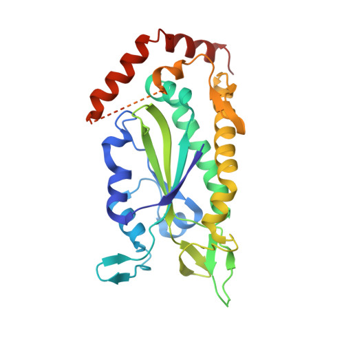
    Conjugative transfer of plasmid DNA via close cell-cell junctions is the main route by which antibiotic resistance genes spread between bacterial strains. Relaxases are essential for conjugative transfer and act by cleaving DNA strands and forming covalent phosphotyrosine linkages. Based on data indicating that multityrosine relaxase enzymes can accommodate two phosphotyrosine intermediates within their divalent metal-containing active sites, we hypothesized that bisphosphonates would inhibit relaxase activity and conjugative DNA transfer. We identified bisphosphonates that are nanomolar inhibitors of the F plasmid conjugative relaxase in vitro. Furthermore, we used cell-based assays to demonstrate that these compounds are highly effective at preventing DNA transfer and at selectively killing cells harboring conjugative plasmids. Two potent inhibitors, clodronate and etidronate, are already clinically approved to treat bone loss. Thus, the inhibition of conjugative relaxases is a potentially novel antimicrobial approach, one that selectively targets bacteria capable of transferring antibiotic resistance and generating multidrug resistant strains.


  • Organizational Affiliation

    Department of Chemistry, Lineberger Comprehensive Cancer Center, University of North Carolina, Chapel Hill, NC 27599-3290, USA.


Macromolecules
Find similar proteins by:  (by identity cutoff)  |  3D Structure
Entity ID: 1
MoleculeChains Sequence LengthOrganismDetailsImage
Protein traI
A, B
301Escherichia coliMutation(s): 1 
Gene Names: traI
EC: 3.6.1
UniProt
Find proteins for P14565 (Escherichia coli (strain K12))
Explore P14565 
Go to UniProtKB:  P14565
Entity Groups  
Sequence Clusters30% Identity50% Identity70% Identity90% Identity95% Identity100% Identity
UniProt GroupP14565
Sequence Annotations
Expand
  • Reference Sequence
Experimental Data & Validation

Experimental Data

  • Method: X-RAY DIFFRACTION
  • Resolution: 2.42 Å
  • R-Value Free: 0.270 
  • R-Value Work: 0.211 
  • R-Value Observed: 0.211 
  • Space Group: P 21 21 21
Unit Cell:
Length ( Å )Angle ( ˚ )
a = 44.888α = 90
b = 88.259β = 90
c = 127.293γ = 90
Software Package:
Software NamePurpose
CNSrefinement
HKL-2000data collection
HKL-2000data reduction
HKL-2000data scaling
MOLREPphasing

Structure Validation

View Full Validation Report


Ligand Structure Quality Assessment 


Entry History 

Deposition Data

Revision History  (Full details and data files)

  • Version 1.0: 2008-05-20
    Type: Initial release
  • Version 1.1: 2011-07-13
    Changes: Version format compliance
  • Version 1.2: 2021-10-20
    Changes: Database references, Derived calculations
  • Version 1.3: 2023-08-30
    Changes: Data collection, Refinement description