2PGB | pdb_00002pgb

Inhibitor-free human thrombin mutant C191A-C220A


Experimental Data Snapshot

  • Method: X-RAY DIFFRACTION
  • Resolution: 1.54 Å
  • R-Value Free: 
    0.217 (Depositor), 0.215 (DCC) 
  • R-Value Work: 
    0.183 (Depositor), 0.184 (DCC) 
  • R-Value Observed: 
    0.183 (Depositor) 

Starting Model: experimental
View more details

wwPDB Validation 3D Report Full Report

Validation slider image for 2PGB

Ligand Structure Quality Assessment 


This is version 1.6 of the entry. See complete history

Literature

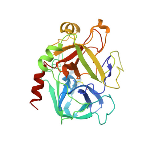
Important role of the cys-191 cys-220 disulfide bond in thrombin function and allostery

Bush-Pelc, L.A.Marino, F.Chen, Z.Pineda, A.O.Mathews, F.S.Di Cera, E.

(2007) J Biological Chem 282: 27165-27170

  • DOI: https://doi.org/10.1074/jbc.M703202200
  • Primary Citation Related Structures: 
    2PGB, 2PGQ

  • PubMed Abstract: 

    Little is known on the role of disulfide bonds in the catalytic domain of serine proteases. The Cys-191-Cys-220 disulfide bond is located between the 190 strand leading to the oxyanion hole and the 220-loop that contributes to the architecture of the primary specificity pocket and the Na+ binding site in allosteric proteases. Removal of this bond in thrombin produces an approximately 100-fold loss of activity toward several chromogenic and natural substrates carrying Arg or Lys at P1. Na+ activation is compromised, and no fluorescence change can be detected in response to Na+ binding. A 1.54-A resolution structure of the C191A/C220A mutant in the free form reveals a conformation similar to the Na+-free slow form of wild type. The lack of disulfide bond exposes the side chain of Asp-189 to solvent, flips the backbone O atom of Gly-219, and generates disorder in portions of the 186 and 220 loops defining the Na+ site. This conformation, featuring perturbation of the Na+ site but with the active site accessible to substrate, offers a possible representation of the recently identified E* form of thrombin. Disorder in the 186 and 220 loops and the flip of Gly-219 are corrected by the active site inhibitor H-D-Phe-Pro-Arg-CH(2)Cl, as revealed by the 1.8-A resolution structure of the complex. We conclude that the Cys-191-Cys-220 disulfide bond confers stability to the primary specificity pocket by shielding Asp-189 from the solvent and orients the backbone O atom of Gly-219 for optimal substrate binding. In addition, the disulfide bond stabilizes the 186 and 220 loops that are critical for Na+ binding and activation.


  • Organizational Affiliation
    • Department of Biochemistry and Molecular Biophysics, Washington University School of Medicine, St. Louis, Missouri 63110.

Macromolecule Content 

  • Total Structure Weight: 34.23 kDa 
  • Atom Count: 2,638 
  • Modeled Residue Count: 289 
  • Deposited Residue Count: 295 
  • Unique protein chains: 2

Macromolecules

Find similar proteins by:|  3D Structure
Entity ID: 1
MoleculeChains  Sequence LengthOrganismDetailsImage
Prothrombin36Homo sapiensMutation(s): 0 
Gene Names: F2
EC: 3.4.21.5
UniProt & NIH Common Fund Data Resources
Find proteins for P00734 (Homo sapiens)
Explore P00734 
Go to UniProtKB:  P00734
PHAROS:  P00734
GTEx:  ENSG00000180210 
Entity Groups
Sequence Clusters30% Identity50% Identity70% Identity90% Identity95% Identity100% Identity
UniProt GroupP00734
Sequence Annotations
Expand
Reference Sequence
Find similar proteins by:|  3D Structure
Entity ID: 2
MoleculeChains  Sequence LengthOrganismDetailsImage
Prothrombin259Homo sapiensMutation(s): 2 
Gene Names: F2
EC: 3.4.21.5
UniProt & NIH Common Fund Data Resources
Find proteins for P00734 (Homo sapiens)
Explore P00734 
Go to UniProtKB:  P00734
PHAROS:  P00734
GTEx:  ENSG00000180210 
Entity Groups
Sequence Clusters30% Identity50% Identity70% Identity90% Identity95% Identity100% Identity
UniProt GroupP00734
Sequence Annotations
Expand
Reference Sequence

Experimental Data & Validation

Experimental Data

  • Method: X-RAY DIFFRACTION
  • Resolution: 1.54 Å
  • R-Value Free:  0.217 (Depositor), 0.215 (DCC) 
  • R-Value Work:  0.183 (Depositor), 0.184 (DCC) 
  • R-Value Observed: 0.183 (Depositor) 
Space Group: P 21 21 2
Unit Cell:
Length ( Å )Angle ( ˚ )
a = 61.412α = 90
b = 96.597β = 90
c = 48.14γ = 90
Software Package:
Software NamePurpose
CNSrefinement
ADSCdata collection
HKL-2000data reduction
SCALEPACKdata scaling
CNSphasing

Structure Validation

View Full Validation Report


Ligand Structure Quality Assessment 


Entry History 

Deposition Data

Revision History  (Full details and data files)

  • Version 1.0: 2007-07-17
    Type: Initial release
  • Version 1.1: 2008-04-21
    Changes: Version format compliance
  • Version 1.2: 2011-07-13
    Changes: Non-polymer description, Version format compliance
  • Version 1.3: 2020-07-29
    Type: Remediation
    Reason: Carbohydrate remediation
    Changes: Data collection, Derived calculations, Structure summary
  • Version 1.4: 2021-10-20
    Changes: Database references, Structure summary
  • Version 1.5: 2023-08-30
    Changes: Data collection, Refinement description
  • Version 1.6: 2024-10-30
    Changes: Structure summary