2OAY

Crystal structure of latent human C1-inhibitor


Experimental Data Snapshot

  • Method: X-RAY DIFFRACTION
  • Resolution: 2.35 Å
  • R-Value Free: 0.218 
  • R-Value Work: 0.174 
  • R-Value Observed: 0.176 

wwPDB Validation   3D Report Full Report


Ligand Structure Quality Assessment 


This is version 1.5 of the entry. See complete history


Literature

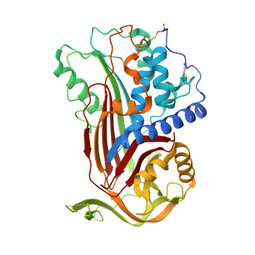
C1 inhibitor serpin domain structure reveals the likely mechanism of heparin potentiation and conformational disease

Beinrohr, L.Harmat, V.Dobo, J.Lorincz, Z.Gal, P.Zavodszky, P.

(2007) J Biol Chem 282: 21100-21109

  • DOI: https://doi.org/10.1074/jbc.M700841200
  • Primary Citation of Related Structures:  
    2OAY

  • PubMed Abstract: 

    C1 inhibitor, a member of the serpin family, is a major down-regulator of inflammatory processes in blood. Genetic deficiency of C1 inhibitor results in hereditary angioedema, a dominantly inheritable, potentially lethal disease. Here we report the first crystal structure of the serpin domain of human C1 inhibitor, representing a previously unreported latent form, which explains functional consequences of several naturally occurring mutations, two of which are discussed in detail. The presented structure displays a novel conformation with a seven-stranded beta-sheet A. The unique conformation of the C-terminal six residues suggests its potential role as a barrier in the active-latent transition. On the basis of surface charge pattern, heparin affinity measurements, and docking of a heparin disaccharide, a heparin binding site is proposed in the contact area of the serpin-proteinase encounter complex. We show how polyanions change the activity of the C1 inhibitor by a novel "sandwich" mechanism, explaining earlier reaction kinetic and mutagenesis studies. These results may help to improve therapeutic C1 inhibitor preparations used in the treatment of hereditary angioedema, organ transplant rejection, and heart attack.


  • Organizational Affiliation

    Institute of Enzymology, Biological Research Center, Hungarian Academy of Sciences, Karolina út 29, H-1113 Budapest, Hungary. lbeinrohr@enzim.hu


Macromolecules
Find similar proteins by:  (by identity cutoff)  |  3D Structure
Entity ID: 1
MoleculeChains Sequence LengthOrganismDetailsImage
Plasma protease C1 inhibitor390Homo sapiensMutation(s): 1 
Gene Names: SERPING1C1INC1NH
UniProt & NIH Common Fund Data Resources
Find proteins for P05155 (Homo sapiens)
Explore P05155 
Go to UniProtKB:  P05155
PHAROS:  P05155
GTEx:  ENSG00000149131 
Entity Groups  
Sequence Clusters30% Identity50% Identity70% Identity90% Identity95% Identity100% Identity
UniProt GroupP05155
Sequence Annotations
Expand
  • Reference Sequence
Experimental Data & Validation

Experimental Data

  • Method: X-RAY DIFFRACTION
  • Resolution: 2.35 Å
  • R-Value Free: 0.218 
  • R-Value Work: 0.174 
  • R-Value Observed: 0.176 
  • Space Group: P 65
Unit Cell:
Length ( Å )Angle ( ˚ )
a = 98.903α = 90
b = 98.903β = 90
c = 94.684γ = 120
Software Package:
Software NamePurpose
MAR345data collection
MOLREPphasing
REFMACrefinement
XDSdata reduction
XSCALEdata scaling

Structure Validation

View Full Validation Report


Ligand Structure Quality Assessment 


Entry History 

Deposition Data

Revision History  (Full details and data files)

  • Version 1.0: 2007-05-01
    Type: Initial release
  • Version 1.1: 2008-05-01
    Changes: Version format compliance
  • Version 1.2: 2011-07-13
    Changes: Advisory, Version format compliance
  • Version 1.3: 2017-10-18
    Changes: Refinement description
  • Version 1.4: 2020-07-29
    Type: Remediation
    Reason: Carbohydrate remediation
    Changes: Data collection, Database references, Derived calculations, Structure summary
  • Version 1.5: 2023-08-30
    Changes: Data collection, Database references, Refinement description, Structure summary