Mutational Analysis of Escherichia coli MoeA: Two Functional Activities Map to the Active Site Cleft.
Nichols, J.D., Xiang, S., Schindelin, H., Rajagopalan, K.V.(2007) Biochemistry 46: 78-86
- PubMed: 17198377 Search on PubMedSearch on PubMed Central
- DOI: https://doi.org/10.1021/bi061551q
- Primary Citation Related Structures:
2NQK, 2NQM, 2NQN, 2NQQ, 2NQR, 2NQS, 2NQU, 2NQV, 2NRO, 2NRP, 2NRS - PubMed Abstract:
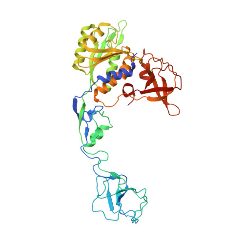
The molybdenum cofactor is ubiquitous in nature, and the pathway for Moco biosynthesis is conserved in all three domains of life. Recent work has helped to illuminate one of the most enigmatic steps in Moco biosynthesis, ligation of metal to molybdopterin (the organic component of the cofactor) to form the active cofactor. In Escherichia coli, the MoeA protein mediates ligation of Mo to molybdopterin while the MogA protein enhances this process in an ATP-dependent manner. The X-ray crystal structures for both proteins have been previously described as well as two essential MogA residues, Asp49 and Asp82. Here we describe a detailed mutational analysis of the MoeA protein. Variants of conserved residues at the putative active site of MoeA were analyzed for a loss of function in two different, previously described assays, one employing moeA- crude extracts and the other utilizing a defined system. Oddly, no correlation was observed between the activity in the two assays. In fact, our results showed a general trend toward an inverse relationship between the activity in each assay. Moco binding studies indicated a strong correlation between a variant's ability to bind Moco and its activity in the purified component assay. Crystal structures of the functionally characterized MoeA variants revealed no major structural changes, indicating that the functional differences observed are not due to disruption of the protein structure. On the basis of these results, two different functional areas were assigned to regions at or near the MoeA active site cleft.
- Department of Biochemistry, Duke University Medical Center, Durham, North Carolina 27710, USA.
Organizational Affiliation: