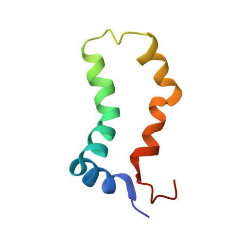
Three-dimensional structure and interaction studies of hepatitis C virus p7 in 1,2-dihexanoyl-sn-glycero-3-phosphocholine by solution nuclear magnetic resonance.
Cook, G.A., Dawson, L.A., Tian, Y., Opella, S.J.(2013) Biochemistry 52: 5295-5303
- PubMed: 23841474 Search on PubMedSearch on PubMed Central
- DOI: https://doi.org/10.1021/bi4006623
- Primary Citation Related Structures:
2MTS - PubMed Abstract:
Hepatitis C virus (HCV) protein p7 plays an important role in the assembly and release of mature virus particles. This small 63-residue membrane protein has been shown to induce channel activity, which may contribute to its functions. p7 is highly conserved throughout the entire range of HCV genotypes, which contributes to making p7 a potential target for antiviral drugs. The secondary structure of p7 from the J4 genotype and the tilt angles of the helices within bilayers have been previously characterized by nuclear magnetic resonance (NMR). Here we describe the three-dimensional structure of p7 in short chain phospholipid (1,2-dihexanoyl-sn-glycero-3-phosphocholine) micelles, which provide a reasonably effective membrane-mimicking environment that is compatible with solution NMR experiments. Using a combination of chemical shifts, residual dipolar couplings, and PREs, we determined the structure of p7 using an implicit membrane potential combining both CS-Rosetta decoys and Xplor-NIH refinement. The final set of structures has a backbone root-mean-square deviation of 2.18 Å. Molecular dynamics simulations in NAMD indicate that several side chain interactions might be taking place and that these could affect the dynamics of the protein. In addition to probing the dynamics of p7, we evaluated several drug-protein and protein-protein interactions. Established channel-blocking compounds such as amantadine, hexamethylene amiloride, and long alkyl chain iminosugar derivatives inhibit the ion channel activity of p7. It has also been shown that the protein interacts with HCV nonstructural protein 2 at the endoplasmic reticulum and that this interaction may be important for the infectivity of the virus. Changes in the chemical shift frequencies of solution NMR spectra identify the residues taking part in these interactions.
- Department of Chemistry and Biochemistry, University of California at San Diego , La Jolla, California 92093, United States.
Organizational Affiliation: