2KUY | pdb_00002kuy

Structure of Glycocin F


Experimental Data Snapshot

  • Method: SOLUTION NMR
  • Conformers Calculated: 20 
  • Conformers Submitted: 12 
  • Selection Criteria: structures with the lowest energy 

wwPDB Validation 3D Report Full Report

Validation slider image for 2KUY

This is version 1.5 of the entry. See complete history

Literature

Structural, dynamic, and chemical characterization of a novel s-glycosylated bacteriocin.

Venugopal, H.Edwards, P.J.Schwalbe, M.Claridge, J.K.Libich, D.S.Stepper, J.Loo, T.Patchett, M.L.Norris, G.E.Pascal, S.M.

(2011) Biochemistry 50: 2748-2755

  • DOI: https://doi.org/10.1021/bi200217u
  • Primary Citation Related Structures: 
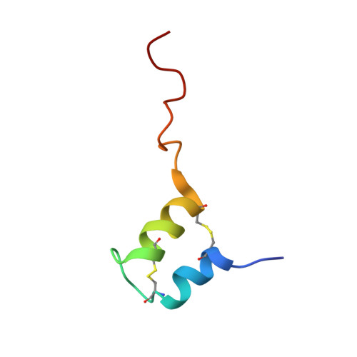
    2KUY

  • PubMed Abstract: 

    Bacteriocins are bacterial peptides with specific activity against competing species. They hold great potential as natural preservatives and for their probiotic effects. We show here nuclear magnetic resonance-based evidence that glycocin F, a 43-amino acid bacteriocin from Lactobacillus plantarum, contains two β-linked N-acetylglucosamine moieties, attached via side chain linkages to a serine via oxygen, and to a cysteine via sulfur. The latter linkage is novel and has helped to establish a new type of post-translational modification, the S-linked sugar. The peptide conformation consists primarily of two α-helices held together by a pair of nested disulfide bonds. The serine-linked sugar is positioned on a short loop sequentially connecting the two helices, while the cysteine-linked sugar presents at the end of a long disordered C-terminal tail. The differing chemical and conformational stabilities of the two N-actetylglucosamine moieties provide clues about the possible mode of action of this bacteriostatic peptide.


  • Organizational Affiliation
    • Institute of Fundamental Sciences, Massey University, Palmerston North, New Zealand. h.venugopal@massey.ac.nz

Macromolecule Content 

  • Total Structure Weight: 5.25 kDa 
  • Atom Count: 361 
  • Modeled Residue Count: 43 
  • Deposited Residue Count: 43 
  • Unique protein chains: 1

Macromolecules

Find similar proteins by:|  3D Structure
Entity ID: 1
MoleculeChains  Sequence LengthOrganismDetailsImage
Prebacteriocin glycocin F43Lactiplantibacillus plantarumMutation(s): 0 
Membrane Entity: Yes 
UniProt
Find proteins for E9K9Z1 (Lactiplantibacillus plantarum)
Explore E9K9Z1 
Go to UniProtKB:  E9K9Z1
Entity Groups
Sequence Clusters30% Identity50% Identity70% Identity90% Identity95% Identity100% Identity
UniProt GroupE9K9Z1
Glycosylation
Glycosylation Sites: 2
Sequence Annotations
Expand
Reference Sequence

Small Molecules

Ligands 1 Unique
IDChains Name / Formula / InChI Key2D Diagram3D Interactions
NAG

Query on NAG


Download:Ideal Coordinates CCD File
B [auth A],
C [auth A]
2-acetamido-2-deoxy-beta-D-glucopyranose
C8 H15 N O6
OVRNDRQMDRJTHS-FMDGEEDCSA-N

Experimental Data & Validation

Experimental Data

  • Method: SOLUTION NMR
  • Conformers Calculated: 20 
  • Conformers Submitted: 12 
  • Selection Criteria: structures with the lowest energy 

Structure Validation

View Full Validation Report


Entry History 

Deposition Data

Revision History  (Full details and data files)

  • Version 1.0: 2011-05-04
    Type: Initial release
  • Version 1.1: 2011-07-13
    Changes: Version format compliance
  • Version 1.2: 2020-02-05
    Changes: Data collection, Derived calculations, Other
  • Version 1.3: 2020-07-29
    Type: Remediation
    Reason: Carbohydrate remediation
    Changes: Advisory, Data collection, Derived calculations, Structure summary
  • Version 1.4: 2023-06-14
    Changes: Database references, Other, Structure summary
  • Version 1.5: 2024-11-20
    Changes: Data collection, Database references, Structure summary