2IUT | pdb_00002iut

P. aeruginosa FtsK motor domain, dimeric


Experimental Data Snapshot

  • Method: X-RAY DIFFRACTION
  • Resolution: 2.25 Å
  • R-Value Free: 
    0.267 (Depositor), 0.210 (DCC) 
  • R-Value Work: 
    0.221 (Depositor), 0.210 (DCC) 
  • R-Value Observed: 
    0.221 (Depositor) 

wwPDB Validation   3D Report Full Report


Ligand Structure Quality Assessment 

Created with Raphaël 2.3.0Worse 01 BetterLigand structure goodness of fit to experimental dataBest fitted AGSClick on this verticalbar to view details

This is version 1.2 of the entry. See complete history


Literature

Double-Stranded DNA Translocation: Structure and Mechanism of Hexameric Ftsk

Massey, T.H.Mercogliano, C.P.Yates, J.Sherratt, D.J.Lowe, J.

(2006) Mol Cell 23: 457

  • DOI: https://doi.org/10.1016/j.molcel.2006.06.019
  • Primary Citation of Related Structures:  
    2IUS, 2IUT, 2IUU

  • PubMed Abstract: 

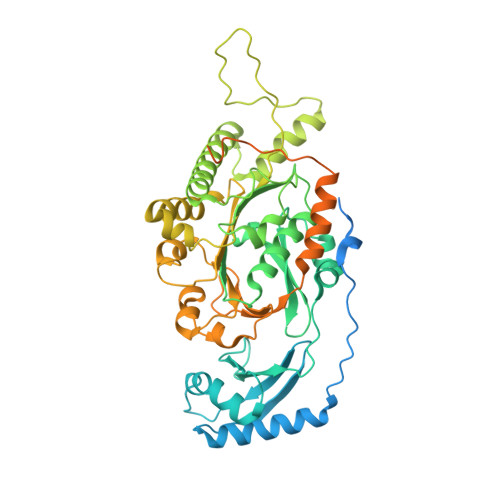
    FtsK is a DNA translocase that coordinates chromosome segregation and cell division in bacteria. In addition to its role as activator of XerCD site-specific recombination, FtsK can translocate double-stranded DNA (dsDNA) rapidly and directionally and reverse direction. We present crystal structures of the FtsK motor domain monomer, showing that it has a RecA-like core, the FtsK hexamer, and also showing that it is a ring with a large central annulus and a dodecamer consisting of two hexamers, head to head. Electron microscopy (EM) demonstrates the DNA-dependent existence of hexamers in solution and shows that duplex DNA passes through the middle of each ring. Comparison of FtsK monomer structures from two different crystal forms highlights a conformational change that we propose is the structural basis for a rotary inchworm mechanism of DNA translocation.


  • Organizational Affiliation

    Division of Molecular Genetics, Department of Biochemistry, University of Oxford, South Parks Road, Oxford OX1 3QU, United Kingdom.


Macromolecules
Find similar proteins by:  (by identity cutoff)  |  3D Structure
Entity ID: 1
MoleculeChains Sequence LengthOrganismDetailsImage
DNA TRANSLOCASE FTSK
A, B
574Pseudomonas aeruginosaMutation(s): 0 
UniProt
Find proteins for Q9I0M3 (Pseudomonas aeruginosa (strain ATCC 15692 / DSM 22644 / CIP 104116 / JCM 14847 / LMG 12228 / 1C / PRS 101 / PAO1))
Explore Q9I0M3 
Go to UniProtKB:  Q9I0M3
Entity Groups  
Sequence Clusters30% Identity50% Identity70% Identity90% Identity95% Identity100% Identity
UniProt GroupQ9I0M3
Sequence Annotations
Expand
  • Reference Sequence
Experimental Data & Validation

Experimental Data

  • Method: X-RAY DIFFRACTION
  • Resolution: 2.25 Å
  • R-Value Free:  0.267 (Depositor), 0.210 (DCC) 
  • R-Value Work:  0.221 (Depositor), 0.210 (DCC) 
  • R-Value Observed: 0.221 (Depositor) 
Space Group: P 1
Unit Cell:
Length ( Å )Angle ( ˚ )
a = 46.168α = 94.57
b = 57.431β = 93.8
c = 98.057γ = 107.66
Software Package:
Software NamePurpose
CNSrefinement
MOSFLMdata reduction
SCALAdata scaling
PHASERphasing

Structure Validation

View Full Validation Report


Ligand Structure Quality Assessment 

Created with Raphaël 2.3.0Worse 01 BetterLigand structure goodness of fit to experimental dataBest fitted AGSClick on this verticalbar to view details

Entry History 

Deposition Data

Revision History  (Full details and data files)

  • Version 1.0: 2006-08-29
    Type: Initial release
  • Version 1.1: 2013-01-30
    Changes: Atomic model, Derived calculations, Non-polymer description, Other, Version format compliance
  • Version 1.2: 2024-05-08
    Changes: Data collection, Database references, Derived calculations, Other