2HKK | pdb_00002hkk

Carbonic anhydrase activators: Solution and X-ray crystallography for the interaction of andrenaline with various carbonic anhydrase isoforms


Experimental Data Snapshot

  • Method: X-RAY DIFFRACTION
  • Resolution: 1.90 Å
  • R-Value Free: 
    0.233 (Depositor), 0.185 (DCC) 
  • R-Value Work: 
    0.186 (Depositor), 0.190 (DCC) 
  • R-Value Observed: 
    0.188 (Depositor) 

Starting Model: experimental
View more details

wwPDB Validation 3D Report Full Report

Validation slider image for 2HKK

Ligand Structure Quality Assessment 


This is version 1.5 of the entry. See complete history

Literature

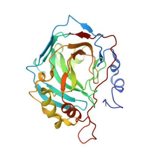
Carbonic anhydrase activators: L-Adrenaline plugs the active site entrance of isozyme II, activating better isoforms I, IV, VA, VII, and XIV.

Temperini, C.Innocenti, A.Scozzafava, A.Mastrolorenzo, A.Supuran, C.T.

(2007) Bioorg Med Chem Lett 17: 628-635

  • DOI: https://doi.org/10.1016/j.bmcl.2006.11.027
  • Primary Citation Related Structures: 
    2HKK

  • PubMed Abstract: 

    The activation of the metalloenzyme carbonic anhydrase (CA, EC 4.2.1.1) with L-adrenaline and histamine has been investigated by kinetic and X-ray crystallographic studies. L-Adrenaline behaves as a potent activator of isozyme CA I (activation constant of 90 nM), being a much weaker activator of isozyme CA II (activation constant of 96 microM). Isoforms CA IV, VA, VII, and XIV were activated by L-adrenaline with K(A)s in the range of 36-63 microM. The X-ray crystal structure of the CA II-L-adrenaline adduct revealed that the activator plugs the entrance of the active site cavity, obstructing it almost completely.


  • Organizational Affiliation
    • Università degli Studi di Firenze, Laboratorio di Chimica Bioinorganica, Rm. 188, Via della Lastruccia 3, I-50019 Sesto Fiorentino (Firenze), Italy.

Macromolecule Content 

  • Total Structure Weight: 29.74 kDa 
  • Atom Count: 2,251 
  • Modeled Residue Count: 256 
  • Deposited Residue Count: 260 
  • Unique protein chains: 1

Macromolecules

Find similar proteins by:|  3D Structure
Entity ID: 1
MoleculeChains  Sequence LengthOrganismDetailsImage
Carbonic anhydrase 2260Homo sapiensMutation(s): 0 
EC: 4.2.1.1 (PDB Primary Data), 4.2.1.69 (UniProt)
UniProt & NIH Common Fund Data Resources
Find proteins for P00918 (Homo sapiens)
Explore P00918 
Go to UniProtKB:  P00918
PHAROS:  P00918
GTEx:  ENSG00000104267 
Entity Groups
Sequence Clusters30% Identity50% Identity70% Identity90% Identity95% Identity100% Identity
UniProt GroupP00918
Sequence Annotations
Expand
Reference Sequence

Experimental Data & Validation

Experimental Data

  • Method: X-RAY DIFFRACTION
  • Resolution: 1.90 Å
  • R-Value Free:  0.233 (Depositor), 0.185 (DCC) 
  • R-Value Work:  0.186 (Depositor), 0.190 (DCC) 
  • R-Value Observed: 0.188 (Depositor) 
Space Group: P 1 21 1
Unit Cell:
Length ( Å )Angle ( ˚ )
a = 42.1α = 90
b = 41.43β = 104.48
c = 72.2γ = 90
Software Package:
Software NamePurpose
AMoREphasing
REFMACrefinement
MOSFLMdata reduction
SCALEPACKdata scaling

Structure Validation

View Full Validation Report


Ligand Structure Quality Assessment 


Entry History 

Deposition Data

Revision History  (Full details and data files)

  • Version 1.0: 2007-05-22
    Type: Initial release
  • Version 1.1: 2007-10-16
    Changes: Version format compliance
  • Version 1.2: 2011-07-13
    Changes: Version format compliance
  • Version 1.3: 2013-03-20
    Changes: Non-polymer description
  • Version 1.4: 2018-04-04
    Changes: Data collection
  • Version 1.5: 2023-08-30
    Changes: Data collection, Database references, Derived calculations, Refinement description