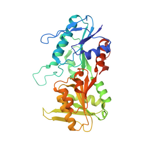
Crystal structure of human phosphoribosylpyrophosphate synthetase 1 reveals a novel allosteric site
Li, S., Lu, Y., Peng, B., Ding, J.(2007) Biochem J 401: 39-47
- PubMed: 16939420 Search on PubMedSearch on PubMed Central
- DOI: https://doi.org/10.1042/BJ20061066
- Primary Citation Related Structures:
2H06, 2H07, 2H08, 2HCR - PubMed Abstract:
PRPP (phosphoribosylpyrophosphate) is an important metabolite essential for nucleotide synthesis and PRS (PRPP synthetase) catalyses synthesis of PRPP from R5P (ribose 5-phosphate) and ATP. The enzymatic activity of PRS is regulated by phosphate ions, divalent metal cations and ADP. In the present study we report the crystal structures of recombinant human PRS1 in complexes with SO4(2-) ions alone and with ATP, Cd2+ and SO4(2-) ions respectively. The AMP moiety of ATP binds at the ATP-binding site, and a Cd2+ ion binds at the active site and in a position to interact with the beta- and gamma-phosphates of ATP. A SO4(2-) ion, an analogue of the activator phosphate, was found to bind at both the R5P-binding site and the allosteric site defined previously. In addi-tion, an extra SO4(2-) binds at a site at the dimer interface between the ATP-binding site and the allosteric site. Binding of this SO4(2-) stabilizes the conformation of the flexible loop at the active site, leading to the formation of the active, open conformation which is essential for binding of ATP and initiation of the catalytic reaction. This is the first time that structural stabilization at the active site caused by binding of an activator has been observed. Structural and biochemical data show that mutations of some residues at this site influence the binding of SO4(2-) and affect the enzymatic activity. The results in the present paper suggest that this new SO4(2-)-binding site is a second allosteric site to regulate the enzymatic activity which might also exist in other eukaryotic PRSs (except plant PRSs of class II), but not in bacterial PRSs.
- State Key Laboratory of Molecular Biology, Institute of Biochemistry and Cell Biology, Shanghai Institutes for Biological Sciences, Chinese Academy of Sciences, 320 Yue-Yang Road, Shanghai 200031, China.
Organizational Affiliation: