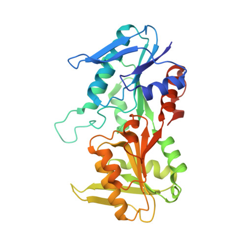
2HCR

crystal structure of human phosphoribosyl pyrophosphate synthetase 1 in complex with AMP(ATP), cadmium and sulfate ion


Experimental Data Snapshot

  • Method: X-RAY DIFFRACTION
  • Resolution: 2.20 Å
  • R-Value Free: 0.254 
  • R-Value Work: 0.205 
  • R-Value Observed: 0.205 

Starting Model: experimental
View more details

wwPDB Validation   3D Report Full Report


Ligand Structure Quality Assessment 


This is version 1.3 of the entry. See complete history


Literature

Crystal structure of human phosphoribosylpyrophosphate synthetase 1 reveals a novel allosteric site

Li, S.Lu, Y.Peng, B.Ding, J.

(2007) Biochem J 401: 39-47

  • DOI: https://doi.org/10.1042/BJ20061066
  • Primary Citation of Related Structures:  
    2H06, 2H07, 2H08, 2HCR

  • PubMed Abstract: 

    PRPP (phosphoribosylpyrophosphate) is an important metabolite essential for nucleotide synthesis and PRS (PRPP synthetase) catalyses synthesis of PRPP from R5P (ribose 5-phosphate) and ATP. The enzymatic activity of PRS is regulated by phosphate ions, divalent metal cations and ADP. In the present study we report the crystal structures of recombinant human PRS1 in complexes with SO4(2-) ions alone and with ATP, Cd2+ and SO4(2-) ions respectively. The AMP moiety of ATP binds at the ATP-binding site, and a Cd2+ ion binds at the active site and in a position to interact with the beta- and gamma-phosphates of ATP. A SO4(2-) ion, an analogue of the activator phosphate, was found to bind at both the R5P-binding site and the allosteric site defined previously. In addi-tion, an extra SO4(2-) binds at a site at the dimer interface between the ATP-binding site and the allosteric site. Binding of this SO4(2-) stabilizes the conformation of the flexible loop at the active site, leading to the formation of the active, open conformation which is essential for binding of ATP and initiation of the catalytic reaction. This is the first time that structural stabilization at the active site caused by binding of an activator has been observed. Structural and biochemical data show that mutations of some residues at this site influence the binding of SO4(2-) and affect the enzymatic activity. The results in the present paper suggest that this new SO4(2-)-binding site is a second allosteric site to regulate the enzymatic activity which might also exist in other eukaryotic PRSs (except plant PRSs of class II), but not in bacterial PRSs.


  • Organizational Affiliation

    State Key Laboratory of Molecular Biology, Institute of Biochemistry and Cell Biology, Shanghai Institutes for Biological Sciences, Chinese Academy of Sciences, 320 Yue-Yang Road, Shanghai 200031, China.


Macromolecules
Find similar proteins by:  (by identity cutoff)  |  3D Structure
Entity ID: 1
MoleculeChains Sequence LengthOrganismDetailsImage
Ribose-phosphate pyrophosphokinase I
A, B
326Homo sapiensMutation(s): 0 
Gene Names: PRPS1
EC: 2.7.6.1
UniProt & NIH Common Fund Data Resources
Find proteins for P60891 (Homo sapiens)
Explore P60891 
Go to UniProtKB:  P60891
PHAROS:  P60891
GTEx:  ENSG00000147224 
Entity Groups  
Sequence Clusters30% Identity50% Identity70% Identity90% Identity95% Identity100% Identity
UniProt GroupP60891
Sequence Annotations
Expand
  • Reference Sequence
Small Molecules
Ligands 3 Unique
IDChains Name / Formula / InChI Key2D Diagram3D Interactions
AMP
Query on AMP

Download Ideal Coordinates CCD File 
G [auth A],
L [auth B]
ADENOSINE MONOPHOSPHATE
C10 H14 N5 O7 P
UDMBCSSLTHHNCD-KQYNXXCUSA-N
CD
Query on CD

Download Ideal Coordinates CCD File 
F [auth A],
K [auth B]
CADMIUM ION
Cd
WLZRMCYVCSSEQC-UHFFFAOYSA-N
SO4
Query on SO4

Download Ideal Coordinates CCD File 
C [auth A]
D [auth A]
E [auth A]
H [auth B]
I [auth B]
C [auth A],
D [auth A],
E [auth A],
H [auth B],
I [auth B],
J [auth B]
SULFATE ION
O4 S
QAOWNCQODCNURD-UHFFFAOYSA-L
Experimental Data & Validation

Experimental Data

  • Method: X-RAY DIFFRACTION
  • Resolution: 2.20 Å
  • R-Value Free: 0.254 
  • R-Value Work: 0.205 
  • R-Value Observed: 0.205 
  • Space Group: H 3
Unit Cell:
Length ( Å )Angle ( ˚ )
a = 170.236α = 90
b = 170.236β = 90
c = 61.82γ = 120
Software Package:
Software NamePurpose
d*TREKdata scaling
CNSrefinement
REFMACrefinement
PDB_EXTRACTdata extraction
CrystalCleardata reduction
CNSphasing

Structure Validation

View Full Validation Report


Ligand Structure Quality Assessment 


Entry History 

Deposition Data

Revision History  (Full details and data files)

  • Version 1.0: 2006-10-24
    Type: Initial release
  • Version 1.1: 2008-04-01
    Changes: Version format compliance
  • Version 1.2: 2011-07-13
    Changes: Derived calculations, Version format compliance
  • Version 1.3: 2023-10-25
    Changes: Data collection, Database references, Derived calculations, Refinement description