2GEH | pdb_00002geh

N-Hydroxyurea, a versatile zinc binding function in the design of metalloenzyme inhibitors


Experimental Data Snapshot

  • Method: X-RAY DIFFRACTION
  • Resolution: 2.00 Å
  • R-Value Free: 
    0.229 (Depositor), 0.198 (DCC) 
  • R-Value Work: 
    0.199 (Depositor), 0.204 (DCC) 
  • R-Value Observed: 
    0.207 (Depositor) 

wwPDB Validation 3D Report Full Report

Validation slider image for 2GEH

This is version 1.4 of the entry. See complete history

Literature

N-Hydroxyurea-A versatile zinc binding function in the design of metalloenzyme inhibitors.

Temperini, C.Innocenti, A.Scozzafava, A.Supuran, C.T.

(2006) Bioorg Med Chem Lett 16: 4316-4320

  • DOI: https://doi.org/10.1016/j.bmcl.2006.05.068
  • Primary Citation Related Structures: 
    2GEH

  • PubMed Abstract: 

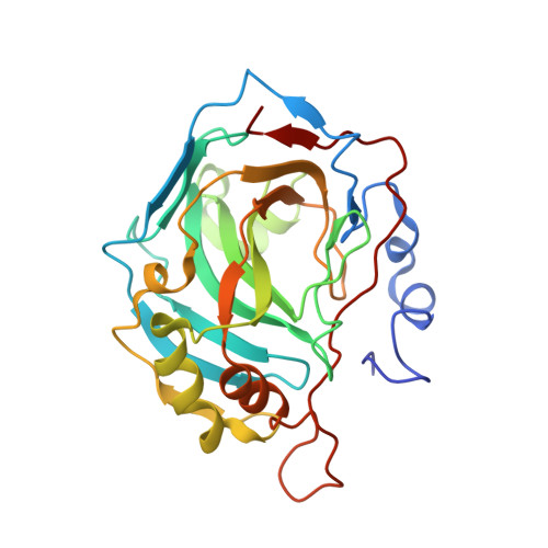
    N-Hydroxyurea binds both to carbonic anhydrase (CA) and to matrix metalloproteinases (MMPs). X-ray crystallography showed N-hydroxyurea to bind in a bidentate mode by means of the oxygen and nitrogen atoms of the NHOH moiety to the Zn(II) ion of CA, participating in a network of hydrogen bonds with a water molecule and Thr199. A derivatized N-hydroxyurea showed low-micromolar affinity for several CAs. This simple zinc binding function may be exploited for obtaining potent metalloenzyme inhibitors, due to its versatility of binding to the metal ion present in the active site of such enzymes.


  • Organizational Affiliation
    • Università degli Studi di Firenze, Laboratorio di Chimica Bioinorganica, Sesto Fiorentino (Firenze), Italy.

Macromolecule Content 

  • Total Structure Weight: 29.83 kDa 
  • Atom Count: 2,233 
  • Modeled Residue Count: 257 
  • Deposited Residue Count: 260 
  • Unique protein chains: 1

Macromolecules

Find similar proteins by:|  3D Structure
Entity ID: 1
MoleculeChains  Sequence LengthOrganismDetailsImage
Carbonic anhydrase 2260Homo sapiensMutation(s): 0 
EC: 4.2.1.1 (PDB Primary Data), 4.2.1.69 (UniProt)
UniProt & NIH Common Fund Data Resources
Find proteins for P00918 (Homo sapiens)
Explore P00918 
Go to UniProtKB:  P00918
PHAROS:  P00918
GTEx:  ENSG00000104267 
Entity Groups
Sequence Clusters30% Identity50% Identity70% Identity90% Identity95% Identity100% Identity
UniProt GroupP00918
Sequence Annotations
Expand
Reference Sequence

Experimental Data & Validation

Experimental Data

  • Method: X-RAY DIFFRACTION
  • Resolution: 2.00 Å
  • R-Value Free:  0.229 (Depositor), 0.198 (DCC) 
  • R-Value Work:  0.199 (Depositor), 0.204 (DCC) 
  • R-Value Observed: 0.207 (Depositor) 
Space Group: P 1 21 1
Unit Cell:
Length ( Å )Angle ( ˚ )
a = 42.201α = 90
b = 41.453β = 104.4
c = 72.268γ = 90
Software Package:
Software NamePurpose
MOSFLMdata reduction
SCALEPACKdata scaling
AMoREphasing
REFMACrefinement

Structure Validation

View Full Validation Report


Entry History 

Deposition Data

Revision History  (Full details and data files)

  • Version 1.0: 2006-06-20
    Type: Initial release
  • Version 1.1: 2008-05-01
    Changes: Version format compliance
  • Version 1.2: 2011-07-13
    Changes: Version format compliance
  • Version 1.3: 2018-04-04
    Changes: Data collection
  • Version 1.4: 2024-02-14
    Changes: Data collection, Database references, Derived calculations