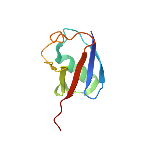
2DEN

Solution Structure of the Ubiquitin-Associated Domain of Human BMSC-UbP and its Complex with Ubiquitin


Experimental Data Snapshot

  • Method: SOLUTION NMR
  • Conformers Calculated: 200 
  • Conformers Submitted: 10 
  • Selection Criteria: structures with the lowest energy 

wwPDB Validation   3D Report Full Report


This is version 1.4 of the entry. See complete history


Literature

Solution structure of the ubiquitin-associated domain of human BMSC-UbP and its complex with ubiquitin

Chang, Y.G.Song, A.X.Gao, Y.G.Shi, Y.H.Lin, X.J.Cao, X.T.Lin, D.H.Hu, H.Y.

(2006) Protein Sci 15: 1248-1259

  • DOI: https://doi.org/10.1110/ps.051995006
  • Primary Citation of Related Structures:  
    2CWB, 2DEN

  • PubMed Abstract: 

    Ubiquitin is an important cellular signal that targets proteins for degradation or regulates their functions. The previously identified BMSC-UbP protein derived from bone marrow stromal cells contains a ubiquitin-associated (UBA) domain at the C terminus that has been implicated in linking cellular processes and the ubiquitin system. Here, we report the solution NMR structure of the UBA domain of human BMSC-UbP protein and its complex with ubiquitin. The structure determination was facilitated by using a solubility-enhancement tag (SET) GB1, immunoglobulin G binding domain 1 of Streptococcal protein G. The results show that BMSC-UbP UBA domain is primarily comprised of three alpha-helices with a hydrophobic patch defined by residues within the C terminus of helix-1, loop-1, and helix-3. The M-G-I motif is similar to the M/L-G-F/Y motifs conserved in most UBA domains. Chemical shift perturbation study revealed that the UBA domain binds with the conserved five-stranded beta-sheet of ubiquitin via hydrophobic interactions with the dissociation constant (KD) of approximately 17 microM. The structural model of BMSC-UbP UBA domain complexed with ubiquitin was constructed by chemical shift mapping combined with the program HADDOCK, which is in agreement with the result from mutagenesis studies. In the complex structure, three residues (Met76, Ile78, and Leu99) of BMSC-UbP UBA form a trident anchoring the domain to the hydrophobic concave surface of ubiquitin defined by residues Leu8, Ile44, His68, and Val70. This complex structure may provide clues for BMSC-UbP functions and structural insights into the UBA domains of other ubiquitin-associated proteins that share high sequence homology with BMSC-UbP UBA domain.


  • Organizational Affiliation

    Key Laboratory of Proteomics, Institute of Biochemistry and Cell Biology, Shanghai Institutes for Biological Sciences, Shanghai 200031, China.


Macromolecules
Find similar proteins by:  (by identity cutoff)  |  3D Structure
Entity ID: 1
MoleculeChains Sequence LengthOrganismDetailsImage
Immunoglobulin G-binding protein G,Ubiquitin-like protein 7108Streptococcus sp. 'group GHomo sapiens
This entity is chimeric
Mutation(s): 1 
Gene Names: spgUBL7BMSCUBPSB132
UniProt & NIH Common Fund Data Resources
Find proteins for Q96S82 (Homo sapiens)
Explore Q96S82 
Go to UniProtKB:  Q96S82
PHAROS:  Q96S82
GTEx:  ENSG00000138629 
Entity Groups  
Sequence Clusters30% Identity50% Identity70% Identity90% Identity95% Identity100% Identity
UniProt GroupQ96S82
Sequence Annotations
Expand
  • Reference Sequence
Find similar proteins by:  (by identity cutoff)  |  3D Structure
Entity ID: 2
MoleculeChains Sequence LengthOrganismDetailsImage
Polyubiquitin-B76Homo sapiensMutation(s): 0 
Gene Names: UBB
UniProt & NIH Common Fund Data Resources
Find proteins for P0CG48 (Homo sapiens)
Explore P0CG48 
Go to UniProtKB:  P0CG48
PHAROS:  P0CG48
GTEx:  ENSG00000150991 
Entity Groups  
Sequence Clusters30% Identity50% Identity70% Identity90% Identity95% Identity100% Identity
UniProt GroupP0CG48
Sequence Annotations
Expand
  • Reference Sequence
Experimental Data & Validation

Experimental Data

  • Method: SOLUTION NMR
  • Conformers Calculated: 200 
  • Conformers Submitted: 10 
  • Selection Criteria: structures with the lowest energy 

Structure Validation

View Full Validation Report


Entry History 

Deposition Data

Revision History  (Full details and data files)

  • Version 1.0: 2006-06-13
    Type: Initial release
  • Version 1.1: 2008-04-30
    Changes: Version format compliance
  • Version 1.2: 2011-07-13
    Changes: Version format compliance
  • Version 1.3: 2020-01-15
    Changes: Database references, Derived calculations, Source and taxonomy, Structure summary
  • Version 1.4: 2024-05-01
    Changes: Data collection, Database references