2C6X | pdb_00002c6x

Structure of Bacillus subtilis citrate synthase


Experimental Data Snapshot

  • Method: X-RAY DIFFRACTION
  • Resolution: 3.40 Å
  • R-Value Free: 
    0.316 (Depositor), 0.299 (DCC) 
  • R-Value Work: 
    0.284 (Depositor), 0.269 (DCC) 
  • R-Value Observed: 
    0.284 (Depositor) 

Starting Model: experimental
View more details

wwPDB Validation 3D Report Full Report

Validation slider image for 2C6X

Ligand Structure Quality Assessment 


This is version 1.4 of the entry. See complete history

Literature

Domain Closure in Citrate Synthases Adapted to the Range of Biological Temperatures

Vogl, C.Hough, D.W.Danson, M.J.Crennell, S.J.

To be published.

Macromolecule Content 

  • Total Structure Weight: 166.54 kDa 
  • Atom Count: 11,672 
  • Modeled Residue Count: 1,452 
  • Deposited Residue Count: 1,452 
  • Unique protein chains: 1

Macromolecules

Find similar proteins by:|  3D Structure
Entity ID: 1
MoleculeChains  Sequence LengthOrganismDetailsImage
CITRATE SYNTHASE 1
A, B, C, D
363Bacillus subtilisMutation(s): 0 
EC: 2.3.3.1 (PDB Primary Data), 2.3.3.16 (UniProt)
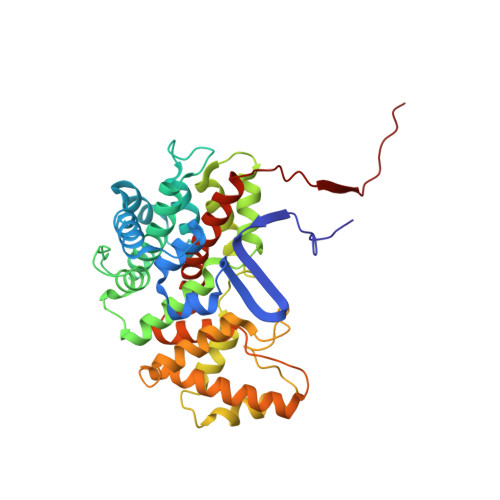
UniProt
Find proteins for P39119 (Bacillus subtilis (strain 168))
Explore P39119 
Go to UniProtKB:  P39119
Entity Groups
Sequence Clusters30% Identity50% Identity70% Identity90% Identity95% Identity100% Identity
UniProt GroupP39119
Sequence Annotations
Expand
Reference Sequence

Small Molecules

Ligands 2 Unique
IDChains Name / Formula / InChI Key2D Diagram3D Interactions
COZ

Query on COZ


Download:Ideal Coordinates CCD File
E [auth A],
G [auth B],
I [auth C],
K [auth D]
COENZYME A
C21 H36 N7 O16 P3 S
RGJOEKWQDUBAIZ-HDCXRZRFSA-N
CIT

Query on CIT


Download:Ideal Coordinates CCD File
F [auth A],
H [auth B],
J [auth C],
L [auth D]
CITRIC ACID
C6 H8 O7
KRKNYBCHXYNGOX-UHFFFAOYSA-N

Experimental Data & Validation

Experimental Data

  • Method: X-RAY DIFFRACTION
  • Resolution: 3.40 Å
  • R-Value Free:  0.316 (Depositor), 0.299 (DCC) 
  • R-Value Work:  0.284 (Depositor), 0.269 (DCC) 
  • R-Value Observed: 0.284 (Depositor) 
Space Group: P 1 21 1
Unit Cell:
Length ( Å )Angle ( ˚ )
a = 53.504α = 90
b = 185.563β = 106.62
c = 82.563γ = 90
Software Package:
Software NamePurpose
CNSrefinement
DENZOdata reduction
SCALEPACKdata scaling
AMoREphasing

Structure Validation

View Full Validation Report


Ligand Structure Quality Assessment 


Entry History 

Deposition Data

Revision History  (Full details and data files)

  • Version 1.0: 2007-06-26
    Type: Initial release
  • Version 1.1: 2011-05-08
    Changes: Version format compliance
  • Version 1.2: 2011-07-13
    Changes: Version format compliance
  • Version 1.3: 2023-12-13
    Changes: Data collection, Database references, Derived calculations, Other, Refinement description
  • Version 1.4: 2025-10-01
    Changes: Advisory, Derived calculations, Structure summary