2BEM

Crystal structure of the Serratia marcescens chitin-binding protein CBP21


Experimental Data Snapshot

  • Method: X-RAY DIFFRACTION
  • Resolution: 1.55 Å
  • R-Value Free: 0.175 
  • R-Value Work: 0.156 
  • R-Value Observed: 0.156 

wwPDB Validation   3D Report Full Report


This is version 1.2 of the entry. See complete history


Literature

Crystal Structure and Binding Properties of the Serratia Marcescens Chitin-Binding Protein Cbp21

Vaaje-Kolstad, G.Houston, D.R.Eijsink, V.G.H.Van Aalten, D.M.F.

(2005) J Biol Chem 280: 11313

  • DOI: https://doi.org/10.1074/jbc.M407175200
  • Primary Citation of Related Structures:  
    2BEM, 2BEN

  • PubMed Abstract: 

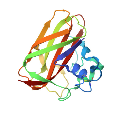
    Chitin proteins are commonly found in bacteria that utilize chitin as a source of energy. CBP21 is a chitin-binding protein from Serratia marcescens, a Gram-negative soil bacterium capable of efficient chitin degradation. When grown on chitin, S. marcescens secretes large amounts of CBP21, along with chitin-degrading enzymes. In an attempt to understand the molecular mechanism of CBP21 action, we have determined its crystal structure at 1.55 angstroms resolution. This is the first structure to be solved of a family 33 carbohydrate-binding module. The structure reveals a "budded" fibronectin type III fold consisting of two beta-sheets, arranged as a beta-sheet sandwich, with a 65-residue "bud" consisting of three short helices, located between beta-strands 1 and 2. Remarkably, conserved aromatic residues that have been suggested previously to play a role in chitin binding were mainly found in the interior of the protein, seemingly incapable of interacting with chitin, whereas the structure revealed a surface patch of highly conserved, mainly hydrophilic residues. The roles of six of these conserved surface-exposed residues (Tyr-54, Glu-55, Glu-60, His-114, Asp-182, and Asn-185) were probed by site-directed mutagenesis and subsequent binding studies. All single point mutations lowered the affinity of CBP21 for beta-chitin, as shown by 3-8-fold increases in the apparent binding constant. Thus, binding of CBP21 to chitin seems to be mediated primarily by conserved, solvent-exposed, polar side chains.


  • Organizational Affiliation

    Department of Chemistry, Biotechnology, and Food Science, Postbox 5003, Agricultural University of Norway, N-1432 As, Norway.


Macromolecules
Find similar proteins by:  (by identity cutoff)  |  3D Structure
Entity ID: 1
MoleculeChains Sequence LengthOrganismDetailsImage
CBP21
A, B, C
170Serratia marcescensMutation(s): 0 
UniProt
Find proteins for O83009 (Serratia marcescens)
Explore O83009 
Go to UniProtKB:  O83009
Entity Groups  
Sequence Clusters30% Identity50% Identity70% Identity90% Identity95% Identity100% Identity
UniProt GroupO83009
Sequence Annotations
Expand
  • Reference Sequence
Small Molecules
Ligands 3 Unique
IDChains Name / Formula / InChI Key2D Diagram3D Interactions
SO4
Query on SO4

Download Ideal Coordinates CCD File 
J [auth A]
Q [auth B]
R [auth B]
S [auth B]
X [auth C]
J [auth A],
Q [auth B],
R [auth B],
S [auth B],
X [auth C],
Y [auth C]
SULFATE ION
O4 S
QAOWNCQODCNURD-UHFFFAOYSA-L
EDO
Query on EDO

Download Ideal Coordinates CCD File 
D [auth A]
E [auth A]
F [auth A]
G [auth A]
H [auth A]
D [auth A],
E [auth A],
F [auth A],
G [auth A],
H [auth A],
I [auth A],
K [auth B],
L [auth B],
M [auth B],
N [auth B],
O [auth B],
P [auth B],
T [auth C],
U [auth C],
V [auth C]
1,2-ETHANEDIOL
C2 H6 O2
LYCAIKOWRPUZTN-UHFFFAOYSA-N
NA
Query on NA

Download Ideal Coordinates CCD File 
W [auth C]SODIUM ION
Na
FKNQFGJONOIPTF-UHFFFAOYSA-N
Experimental Data & Validation

Experimental Data

  • Method: X-RAY DIFFRACTION
  • Resolution: 1.55 Å
  • R-Value Free: 0.175 
  • R-Value Work: 0.156 
  • R-Value Observed: 0.156 
  • Space Group: P 21 21 21
Unit Cell:
Length ( Å )Angle ( ˚ )
a = 51.936α = 90
b = 58.053β = 90
c = 159.829γ = 90
Software Package:
Software NamePurpose
CNSrefinement
DENZOdata reduction
SCALEPACKdata scaling
CNSphasing

Structure Validation

View Full Validation Report


Entry History 

Deposition Data

Revision History  (Full details and data files)

  • Version 1.0: 2004-12-08
    Type: Initial release
  • Version 1.1: 2011-05-08
    Changes: Version format compliance
  • Version 1.2: 2011-07-13
    Changes: Version format compliance